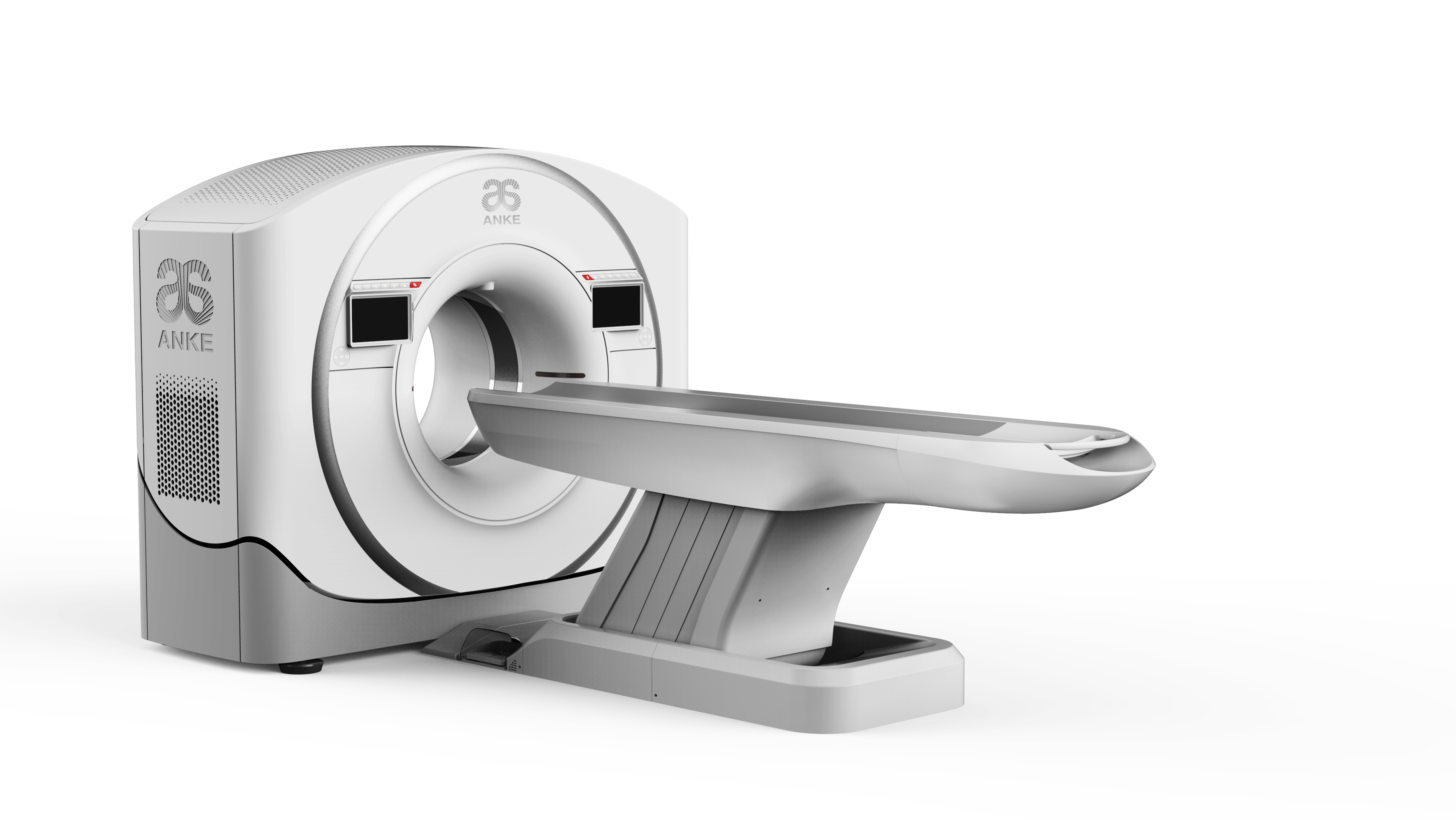
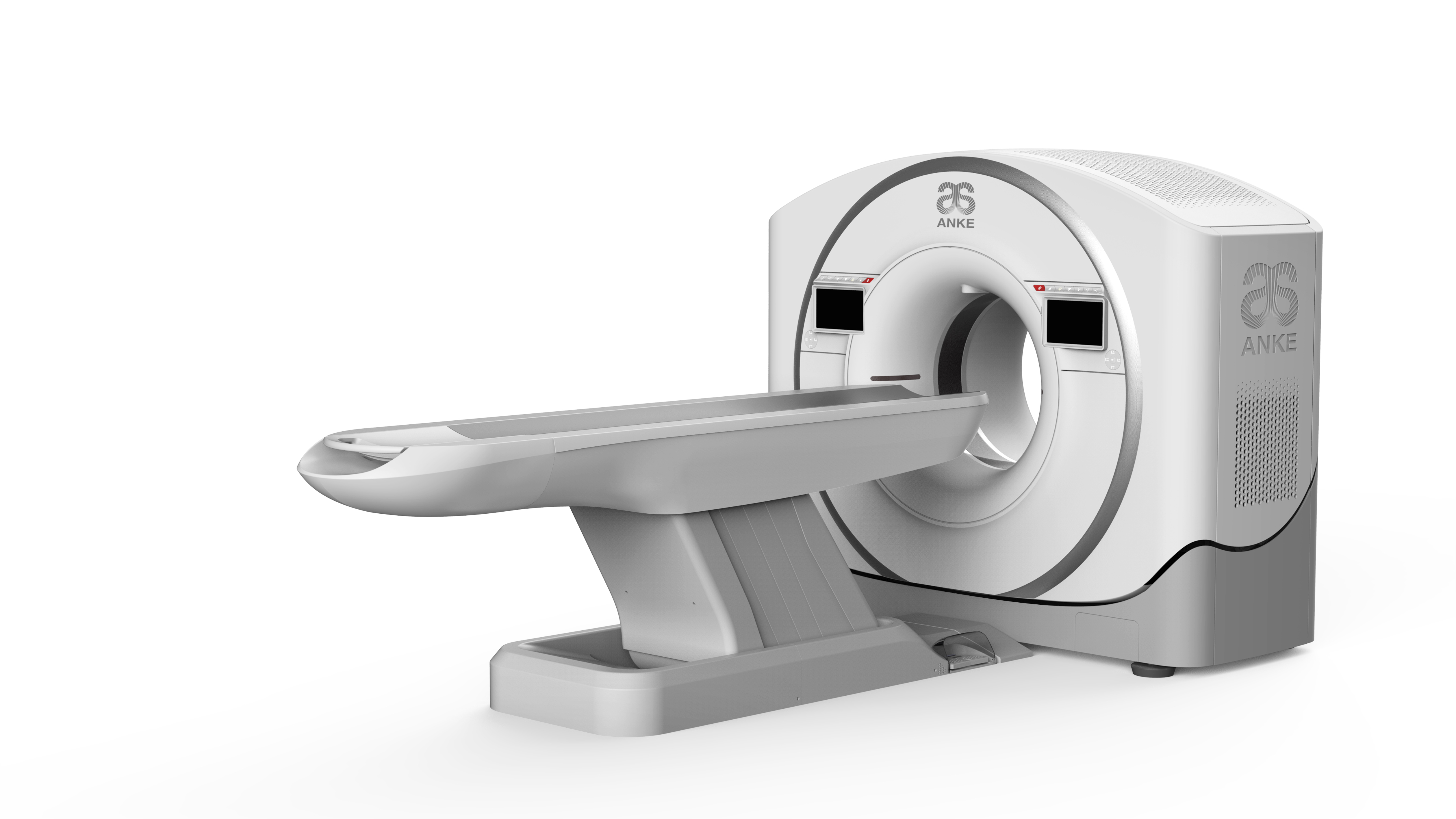
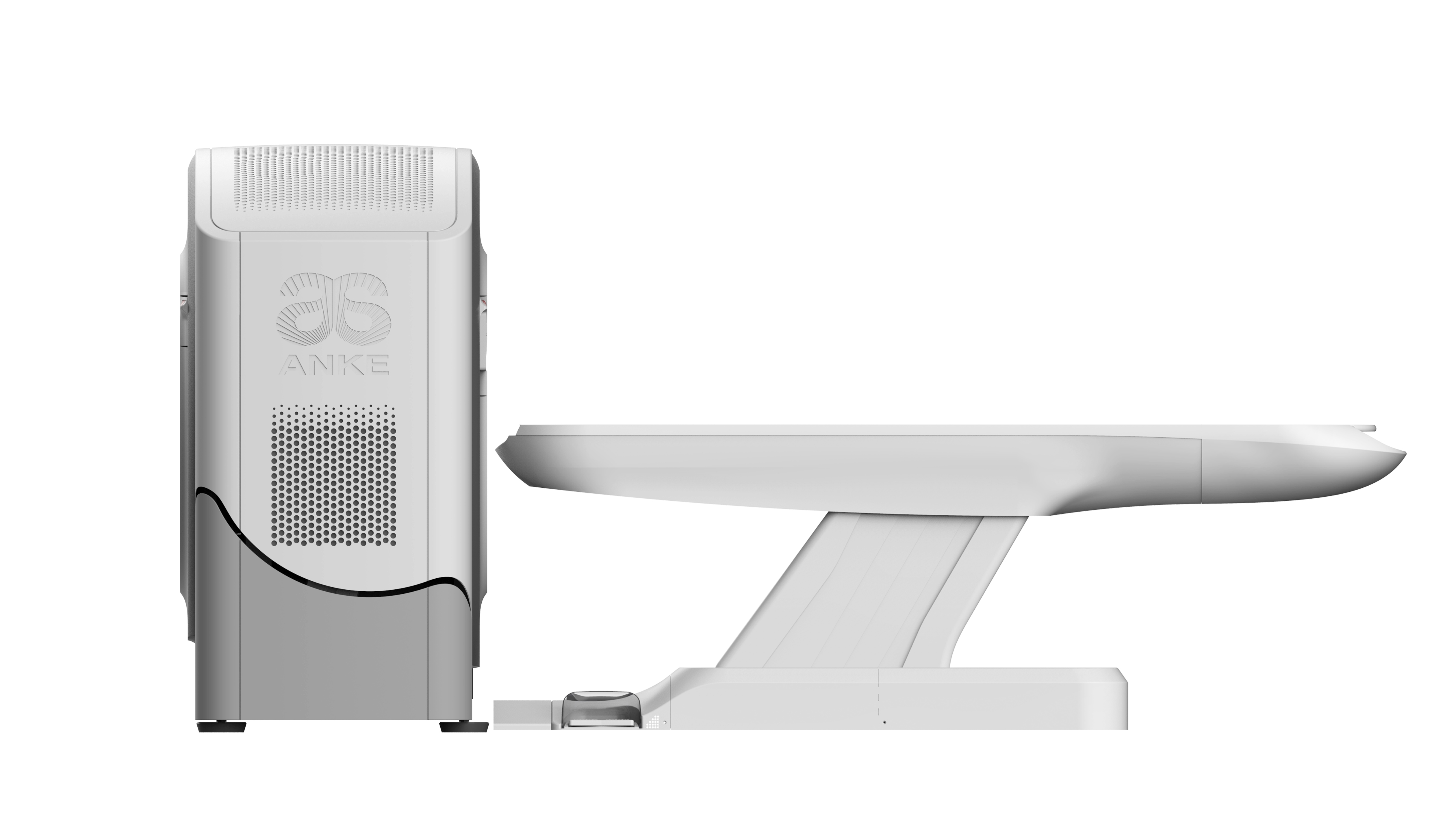
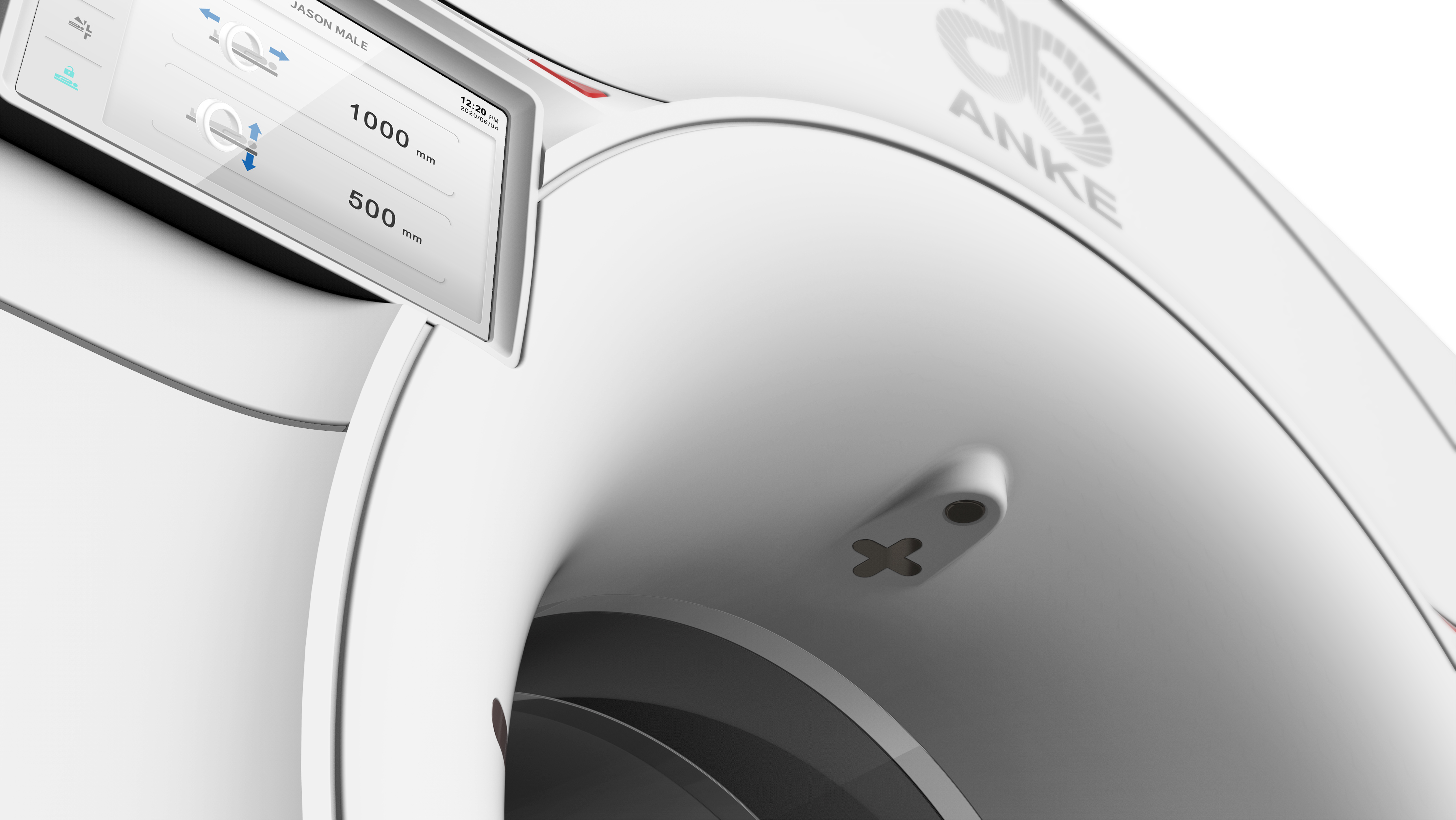
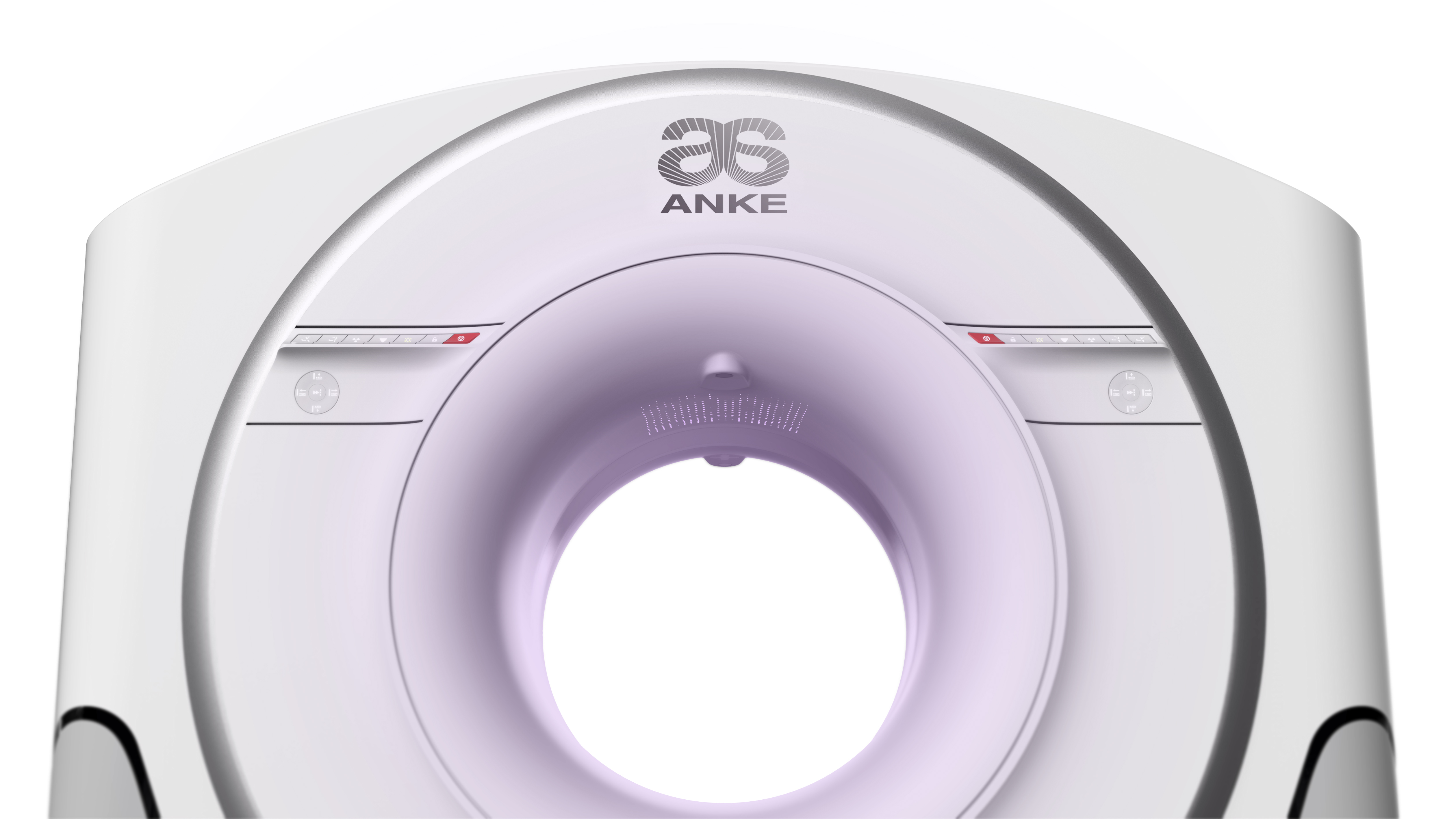
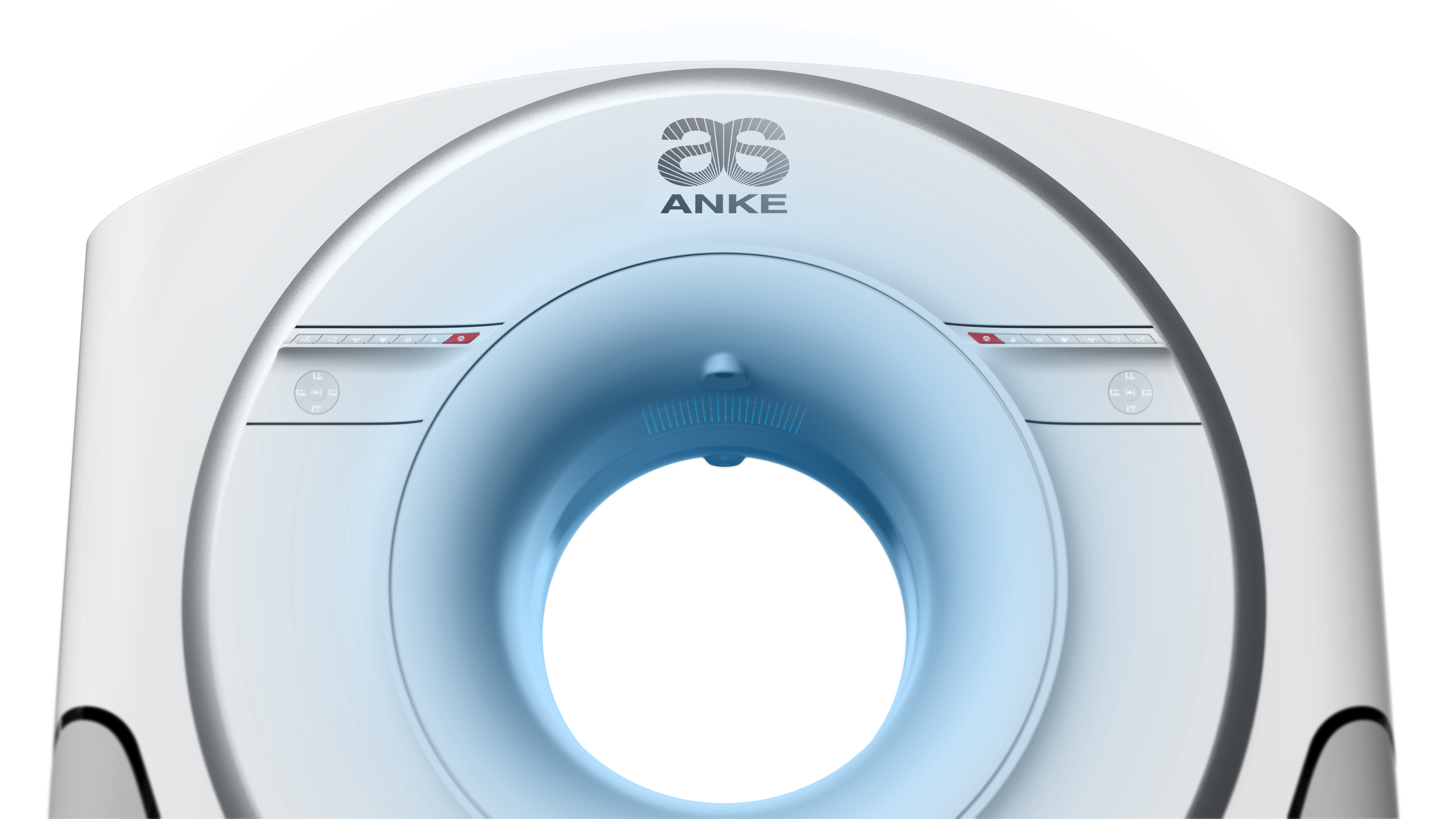
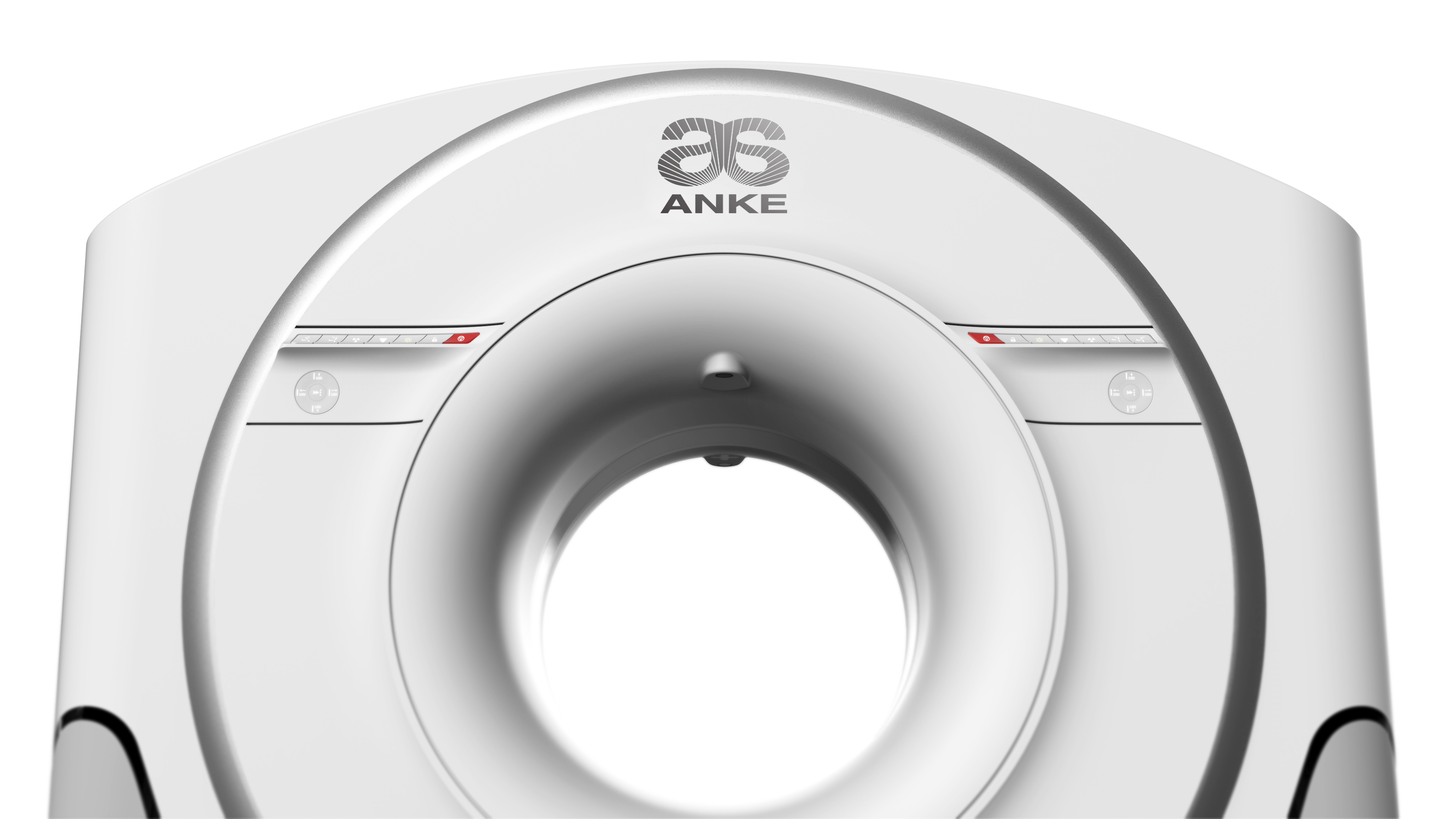
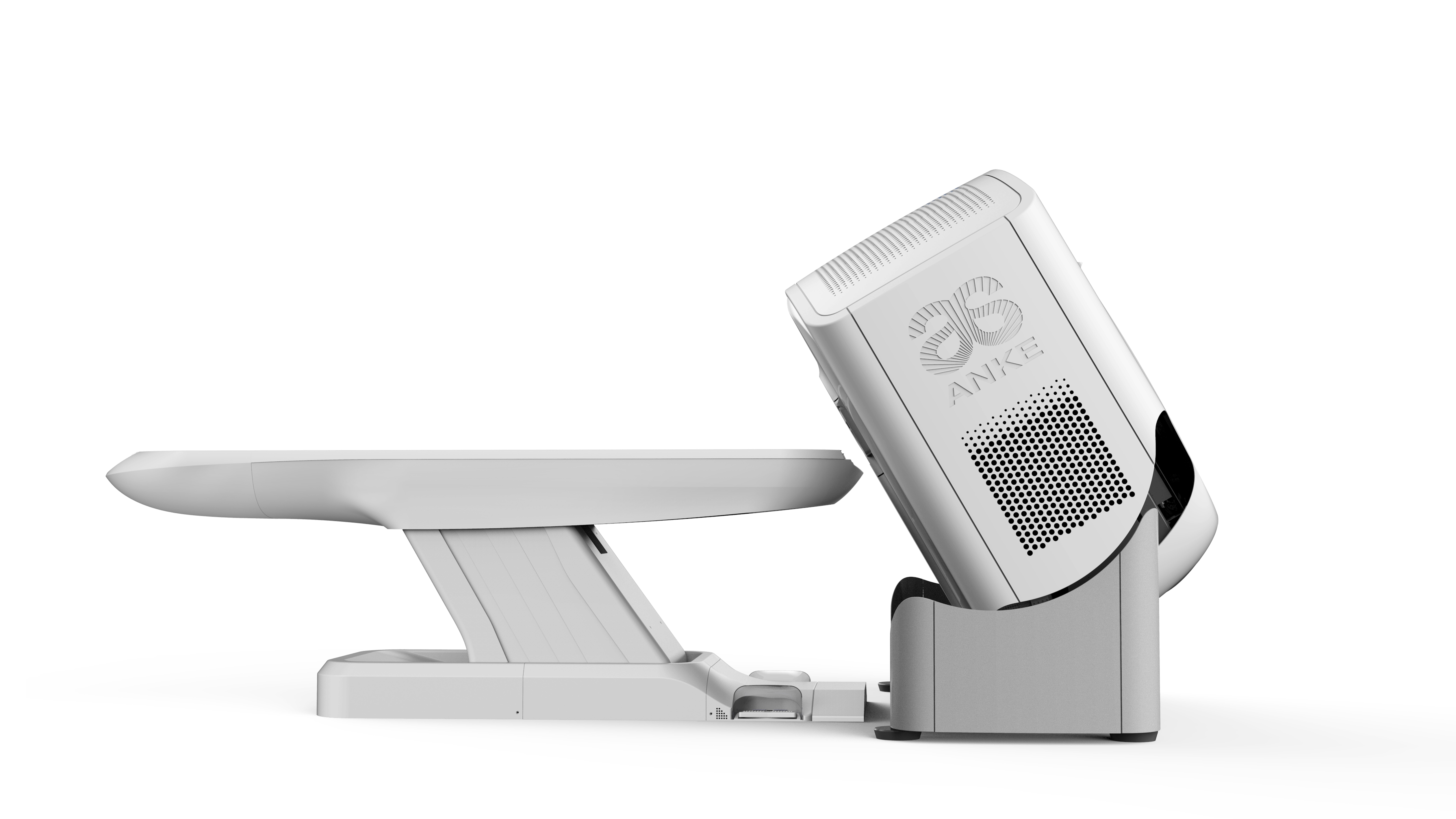
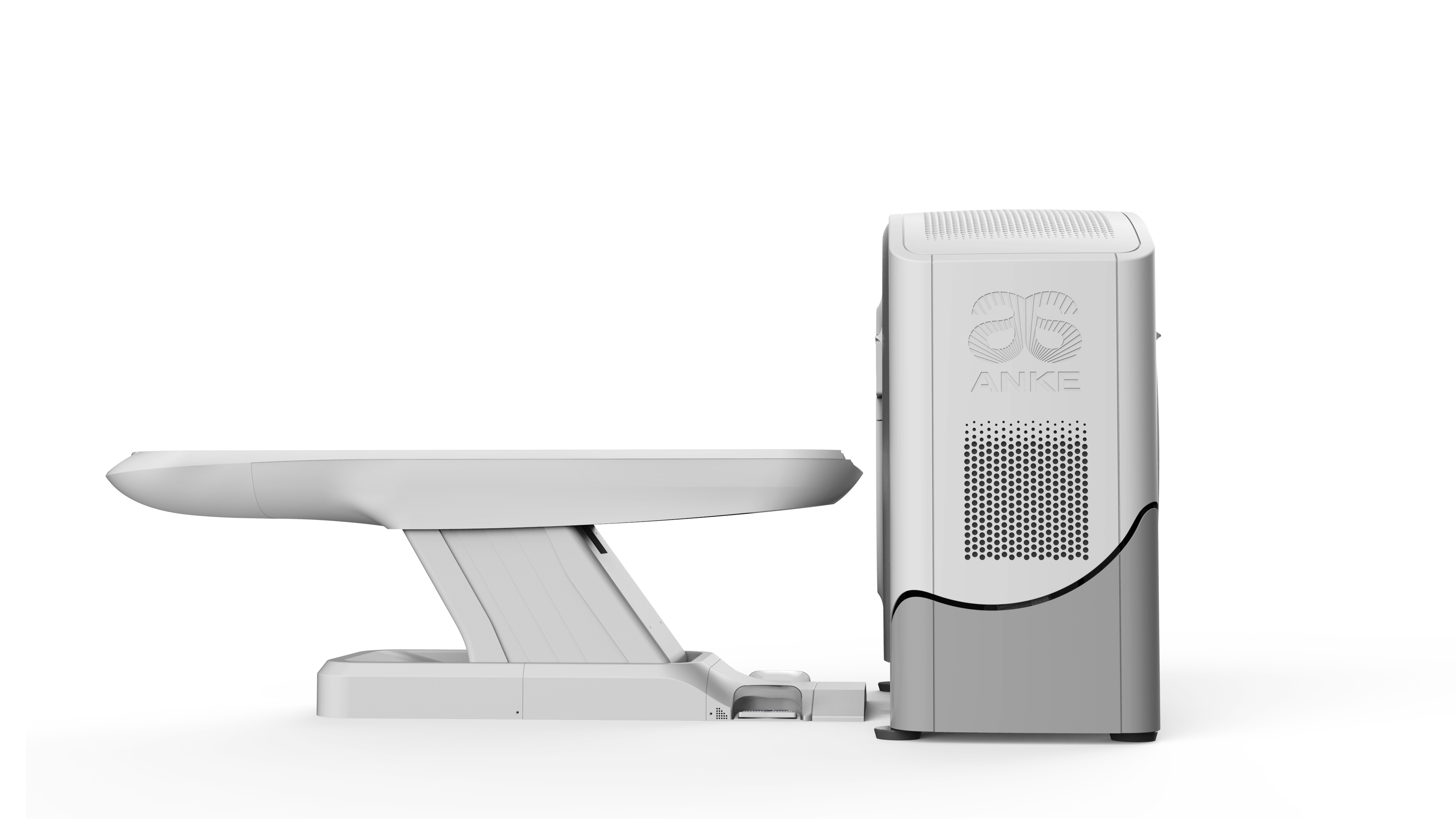
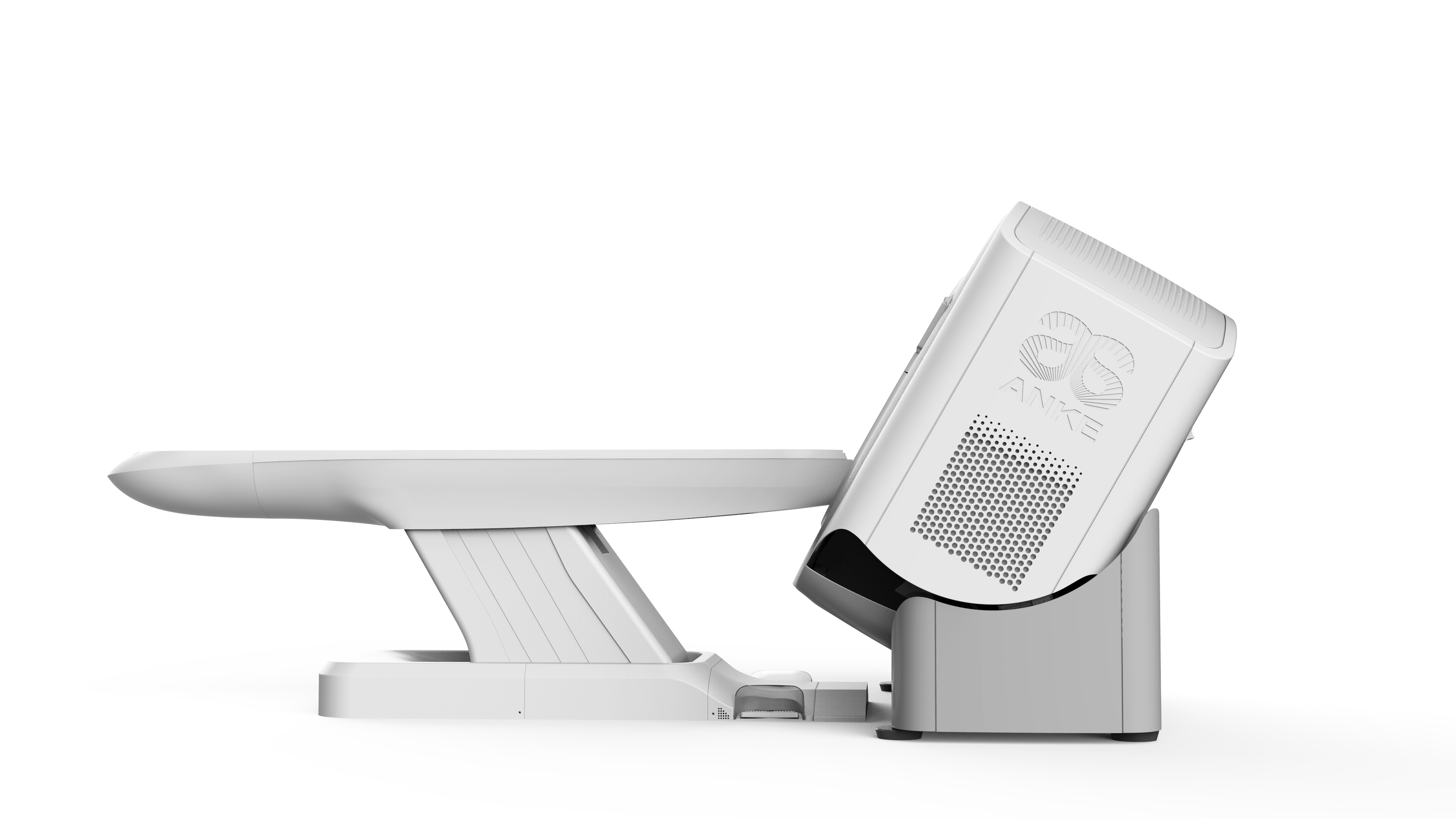
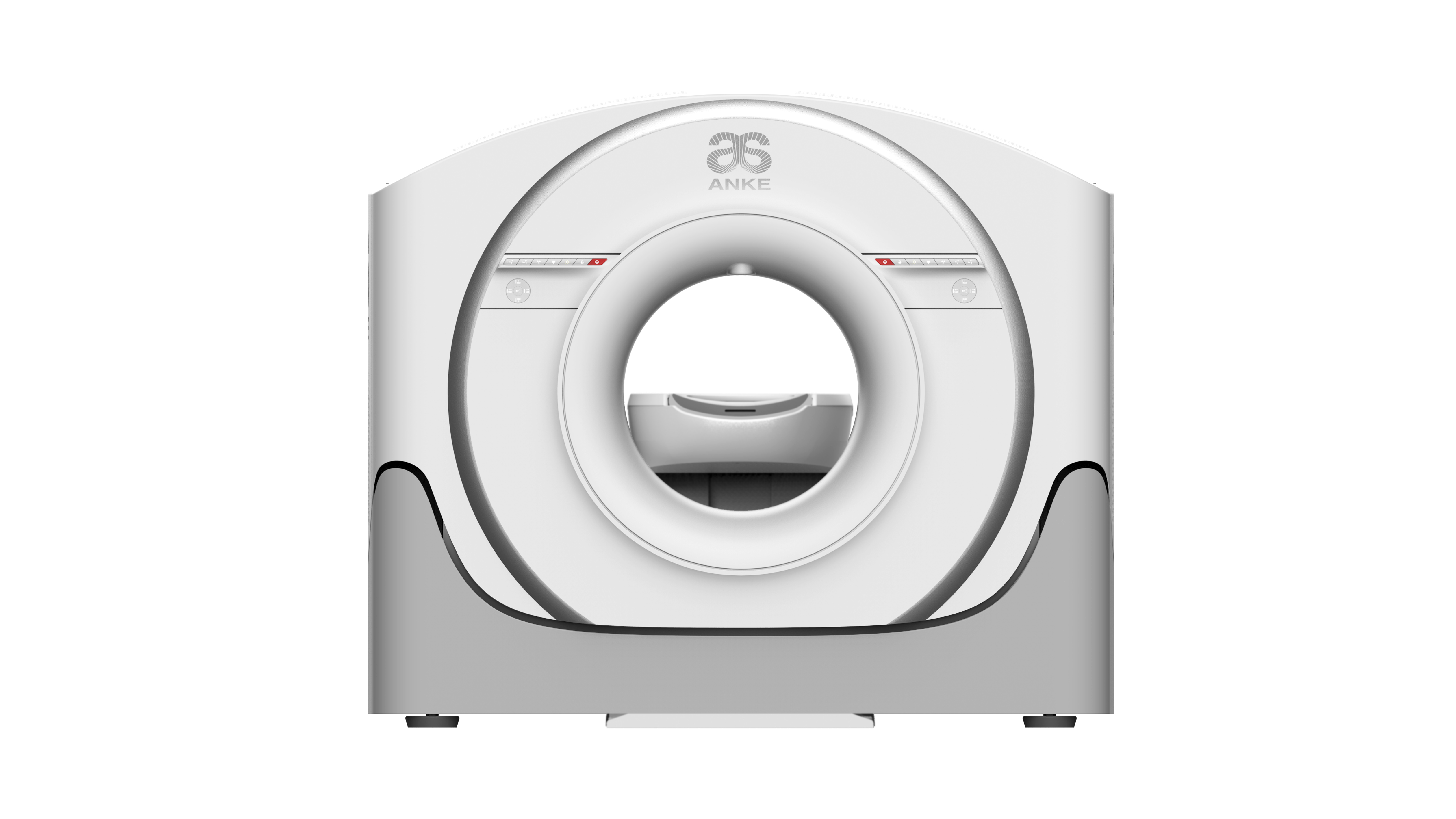
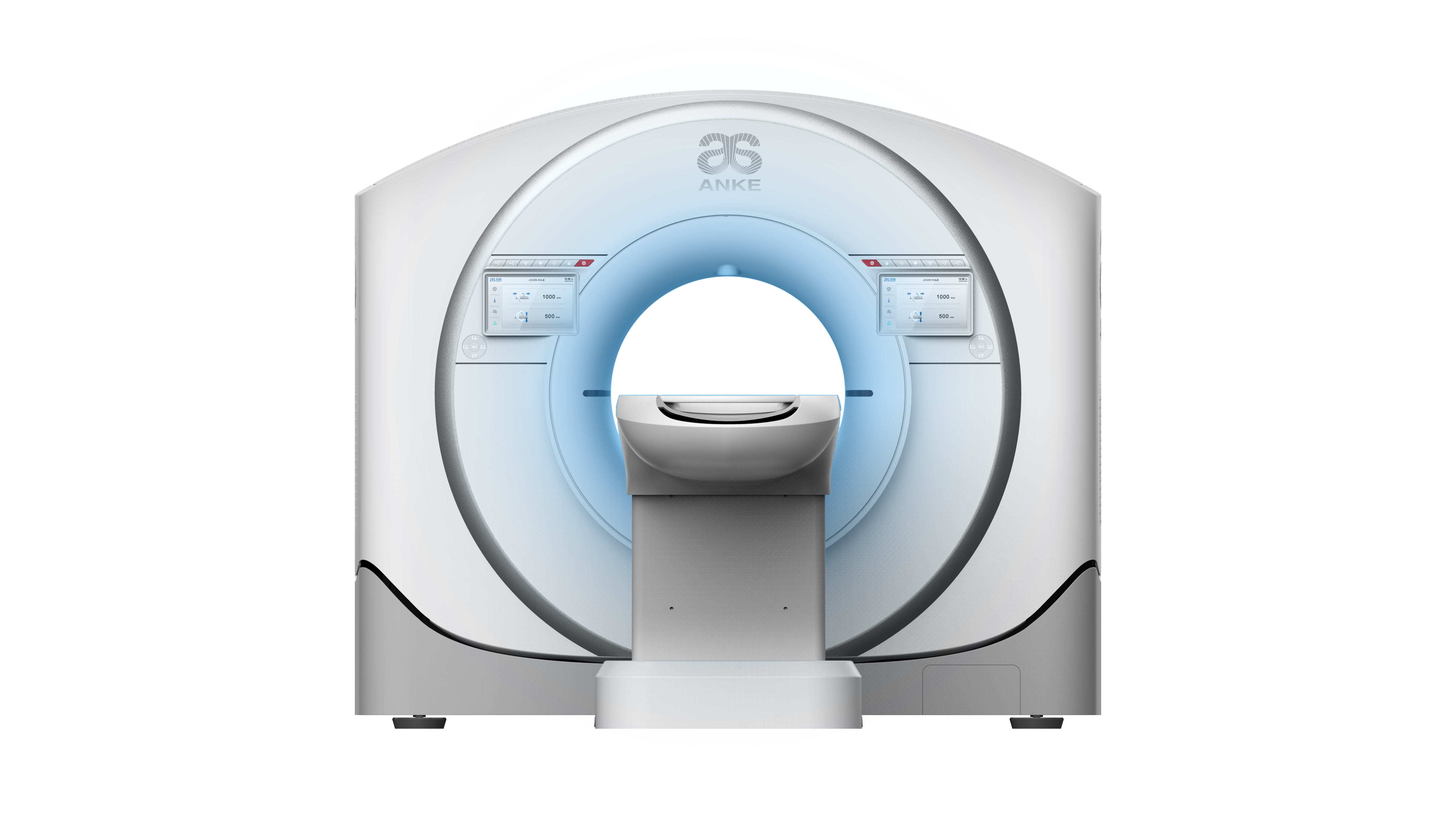
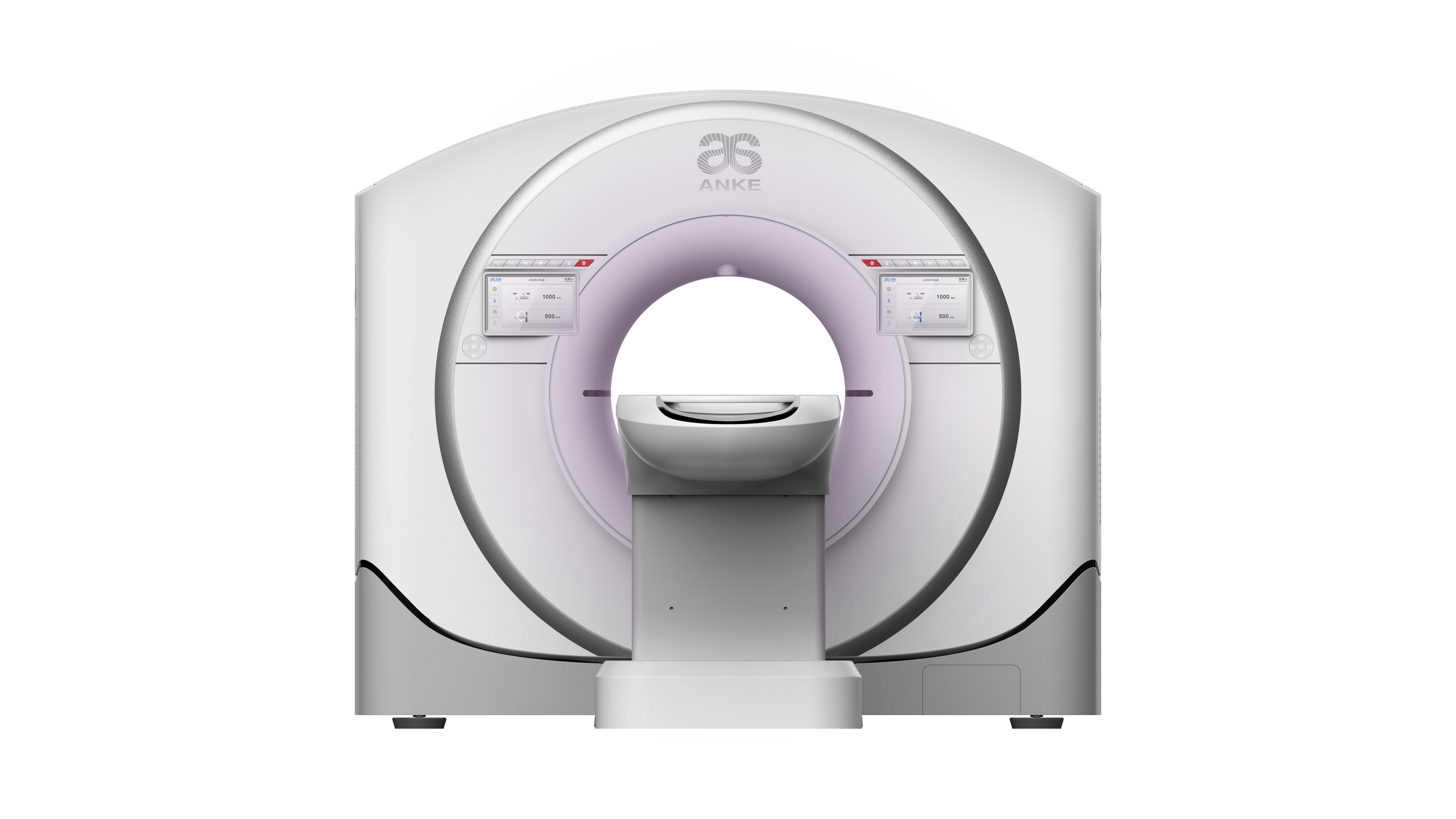
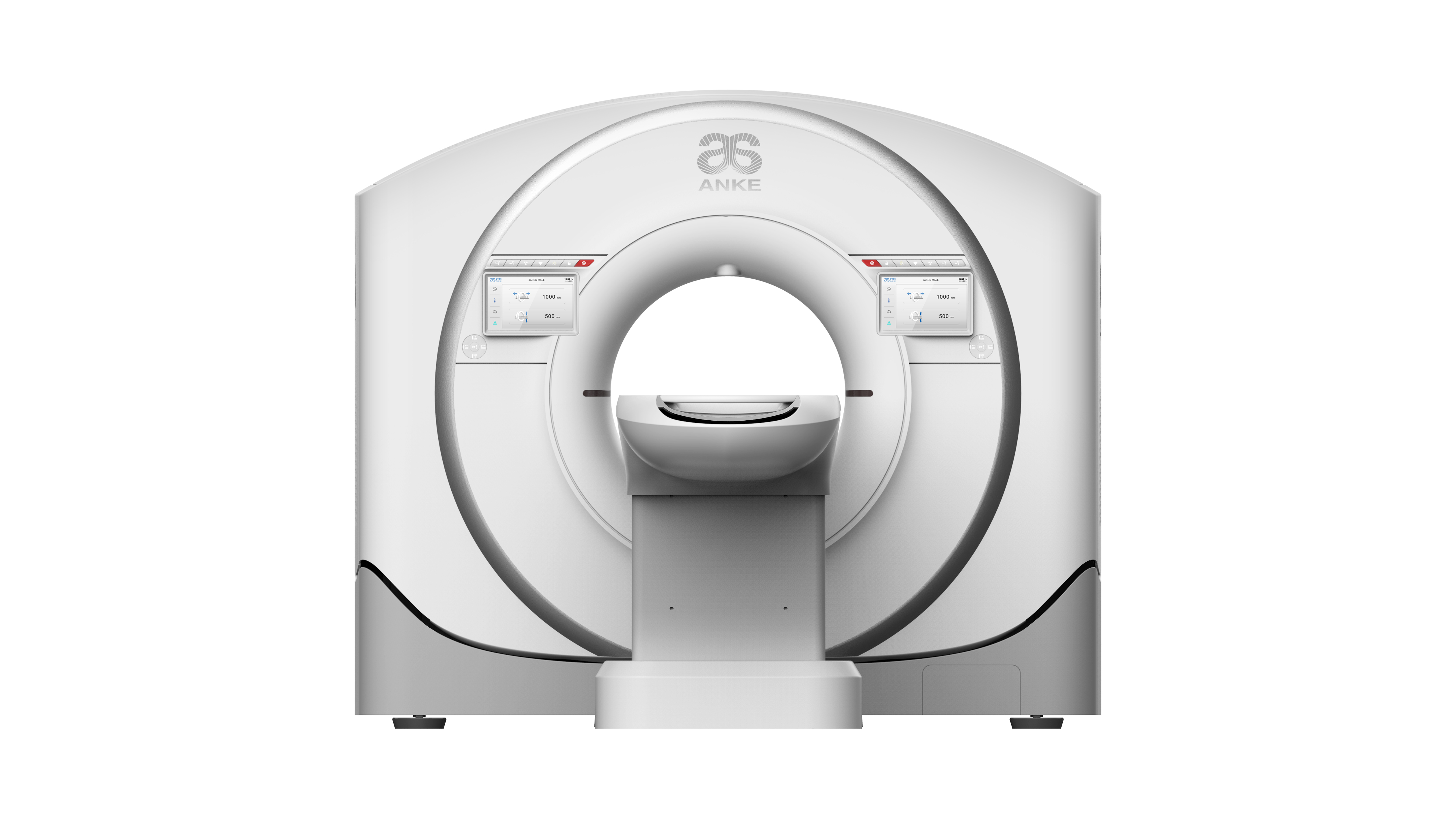
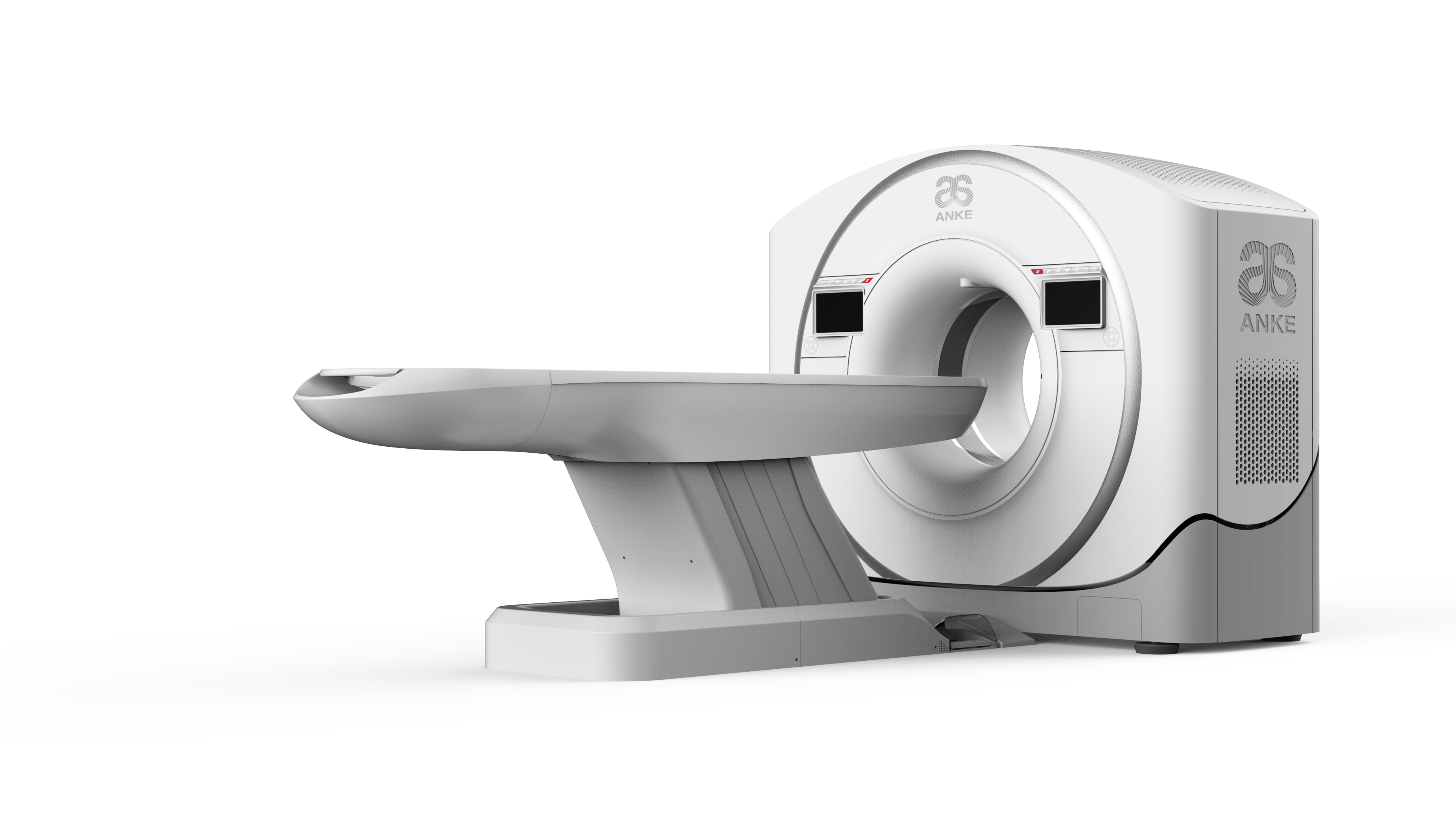
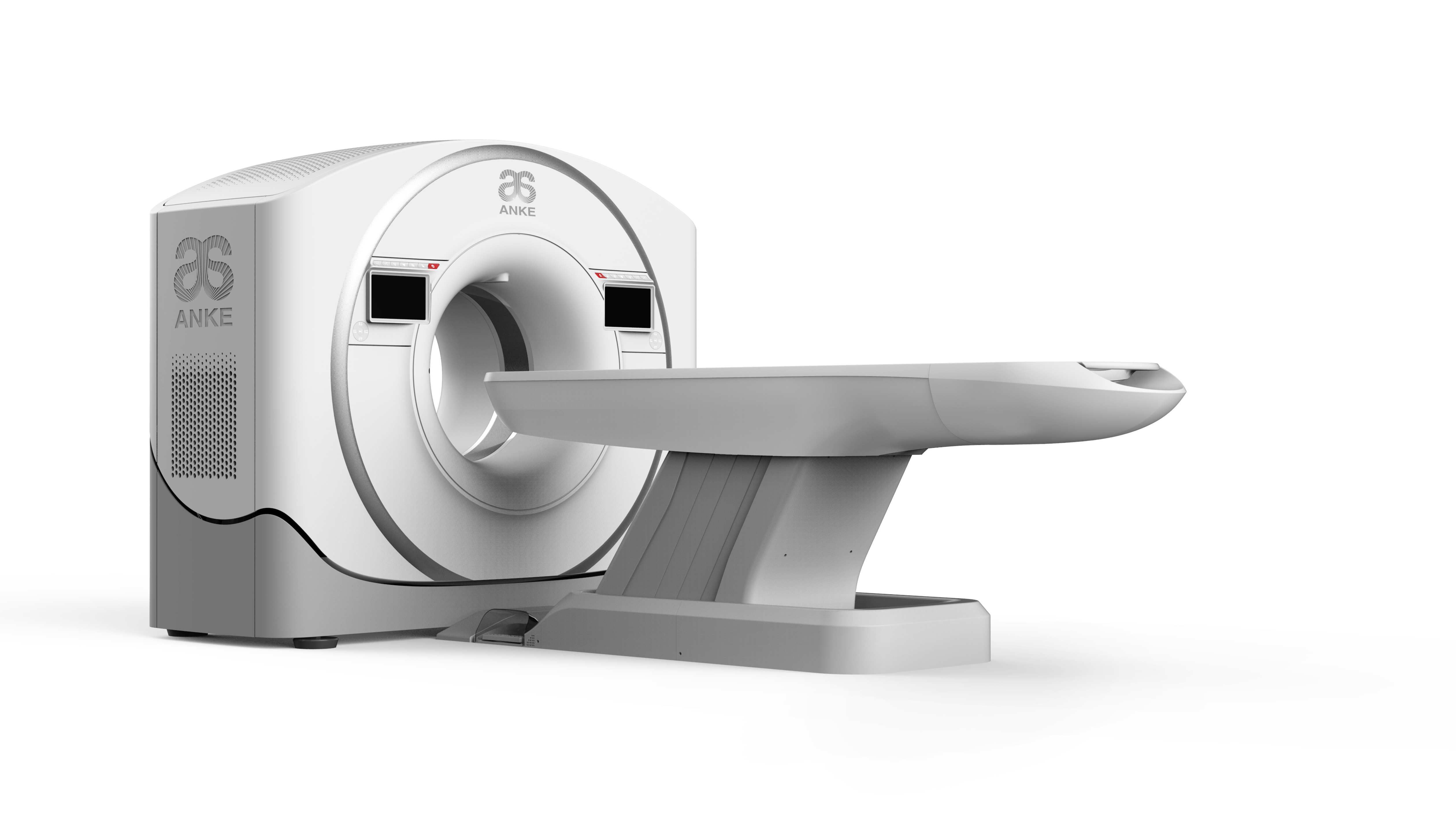
ANATOM 64 Precision adopts ANKE’s self-developed 2-cm OptiWave detector, which is organised and supported by the Ministry of Industry and Information Technology of the People’s Republic of China. It is designed with an isofocal hardware structure, combined with the AccuShape3D stereoscopic anti-scattering grids, so that the distance from the X-rays to the corresponding receiving unit of the detector is the same. In addition of obtaining high quality, high resolution images, it also achieves 2cm wide body coverage in both axial and spiral scanning, which not only improves examination efficiency but also enriches clinical applications.
Field replaceable unit (FRU) design, the key parts be easily disassembled, all are in order to achieve the purpose of simplifying the maintenance operation and reducing the maintenance time. Each detector module can be independent of disassembly and replacement which makes the detector module maintenance and system upgrades completed quickly at the scene. It can reduce the user’s operation and maintenance costs, on the other hand, ensure the equipment with continuous upgrading ability.
Under the same conditions, the service life of the CT detector depends on the decay amplitude of the output signal induced by accumulated exposure dose over time. OptiWave detector has been designed of the optimal structure, even after 10 years of use, the radiation damage caused by signal attenuation is less than 8%. It not only prolongs the service life of the detector but also ensures the users’ maximum return on investment.
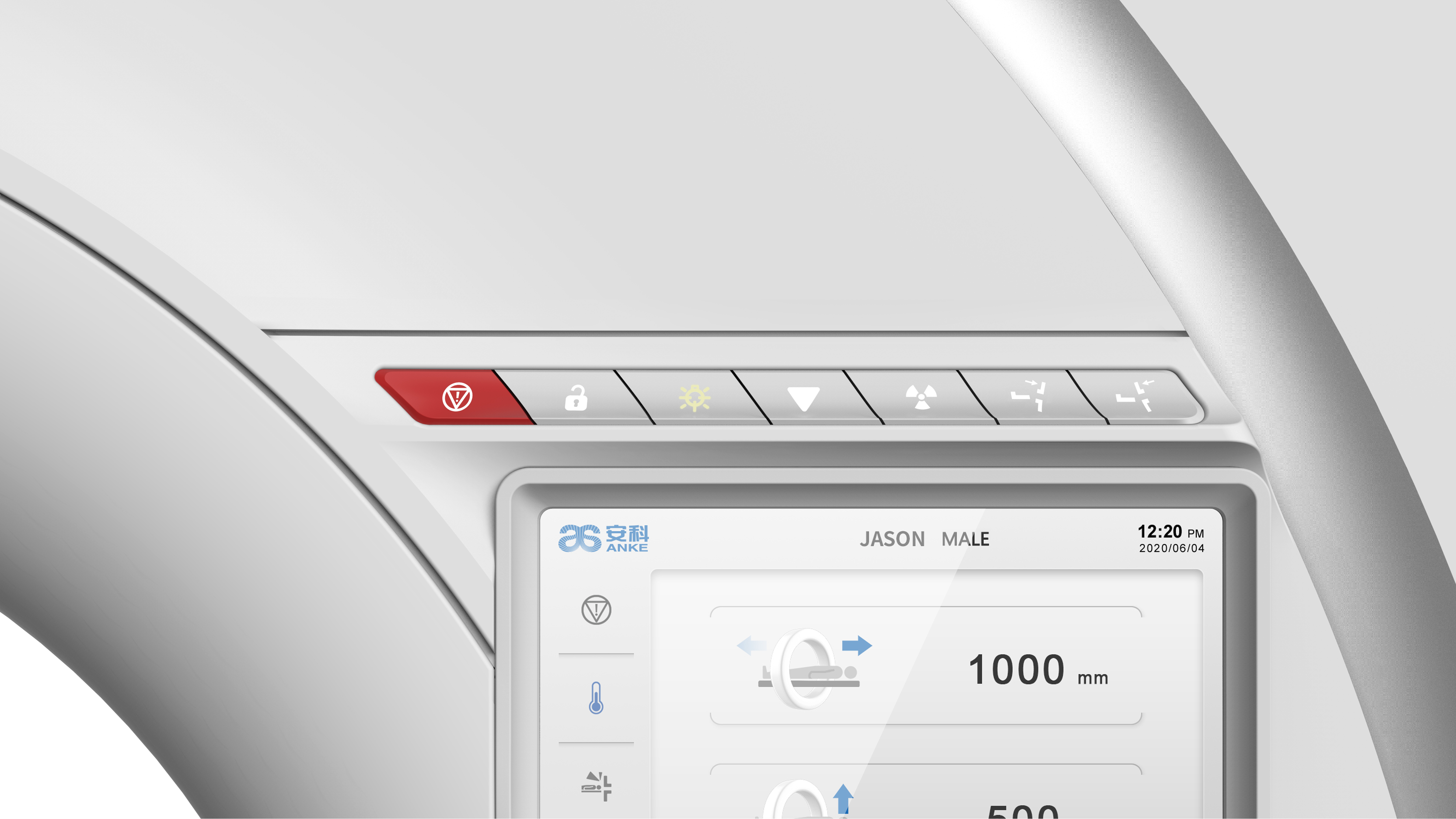
Whith the help of a deep binocular visual perception system-AccuPositioning, deep learning is used to give the device the cognitive ability and behaviour to enable the system to intelligently identify multiple positioning points on the human body and display the scanning site on the intelligent touch screen terminal. As well it enables automatically identify the isocentric position of the proposed scanning site to achieve precise and intelligent patient positioning. Through the application of this technology, it not only substantially improves the accuracy of patient positioning and reduces operational errors, but also better protects patients from the risk of collision and accidental injury. More importantly, the AccuPositioning system also helps the technician to standardise operations and mitigate the increase in radiation dose to the patient’s surface due to inaccurate positioning, while further reducing image noise, reducing artefacts and improving image quality.
ANATOM 64 Precision uses the world leading Admir3D iterative reconstruction technology, through innovative designs of data sampling technology, original data reconstruction technology, post-processing technology and others, Admir3D not only can fully extract the effective data information, but also the data information In the original data field, the corrected projection field and the image field three different data space for multiple recycling, processing, to increase the number of original data reconstruction. That can reduce the X-ray dose and the image noise but Improve the image quality, while access to various parts of high-resolution, low dose of clinical images.
Traditional CT in order to improve the image quality can only be achieved by increasing the dose of X- ray, but at the same time the patient’s radiation dose will also be greatly increased. In order to better solve the problem of image quality and radiation dose, Admir3D accurately constructs and describes the photon characteristics of the signal through a unique mathematical model, iterates it in the original data domain, the corrected projection domain and the image domain. Admir3D processing effect can be performed from 1 to 100 levels, corresponding to the noise reduction requirements.
Admir3D fast reconstruction engine technology
Admir3D iteration technology is based on the original data field, the projection field after correction and image field, which will no doubt greatly increase the system’s computing load. According to this feature, Admir3D platform is equipped with Intel 8-core dual-CPU parallel processor and 64G RAM. Its powerful data processing capability allows the system to mass data processing speed increased nearly 5 times.
Gantry’s built-in camera-“Eagle Eye”allows real-time monitoring of the patient’s status, together with the AI-based AccuClear function, and enables intelligent real time image artifact correction. The novel design of the built-in camera allows for both remote observation of the person being examined and also real-time motion recognition. With AI, correction of motion artefacts can be performed. It is particularly helpful for scanning patients who have involuntary movements, subconscious movements, etc.
Early detection, diagnosis and treatment have been agreed upon for many years, but achieving this is challenging, especially for small lesions (<0.6mm diameter), which are often missed due to the volumetric effect of thicker films. Although sub-millimetre acquisition and reconstruction is now possible with the vast majority of devices, the diagnosis is hampered by the low signal-to-noise ratio of thin-slice images. Based on the above situation, ANKE proposes the AI-based high-resolution thin-slice image reconstruction technology ASR (AI Super Resolution reconstruction), which uses a neural network processing unit to correct and reconstruct the artifacts of the overlapping projection data with artifacts by means of deep learning, which can significantly reduce the overlapping artifacts, and improve the resolution of CT images. By obtaining super thin-slice images with a high resolution of 0.3125mm, you will improve the accuracy of clinical diagnosis.
Spectrum imaging was implemented in 1973 by Hounsfield, the inventor of CT, by using two tube voltages for sequential scanning to distinguish between substances of different atomic number. Alvarez and Macovski, the first to explain the principle of CT dual energy imaging, pointed out that X-rays with mixed energy penetrate the body at the same time and that the Photoelectric and Compton effects produced in different substances can be used for CT Spectrum imaging.
There are two commonly used methods of X-ray energy decomposition, one is based on projection data domain, the other is image data domain. Studies have shown that the former is more accurate and facilitates the removal of artefacts, especially if the data is perfectly matched in time and space.
Currently, there are a number of proven CT energy imaging modalities available on market, including dual-source energy, high & low-kV switching, dual-layer detector and dual-helix/dual-axis.
Among them, dual helix/dual axis energy imaging has certain limitations (limited to sites or organs with autonomous motion) and is often used in low-end CT products; while dual source CT, dual layer detector CT and kV switching energy Spectrum CT can obtain good energy separation effect, but due to the high acquisition and maintenance costs, they cannot be widely used and promoted. In view of this, we have introduced the “Dual-scan Spectrum Imaging” technology based on conventional X-ray generation systems and OptiWave detectors and which is combined with Adose mA modulation technology and encoder assisted projection data alignment to achieve energy separation.
CCTA imaging, as the most important clinical application of post 64-row CT, has always been the most important technical point of interest for users during the procurement and use process. How can the success rate be improved? How can we make the examination more comfortable? How can we meet the needs of patients with high heart rates? How to meet the needs of patients with irregular heart rates? How to achieve low radiation dose and low contrast dosage for coronary imaging? Can energy spectrum imaging be applied to CCTA imaging? How can coronary artery analysis and diagnosis be performed quickly? How can we provide more accurate information to clinicians? …All these questions are the difficulties and pain points encountered by the majority of medical technicians in actual clinical work. In order to effectively solve these objective and practical problems, ANKE uses advanced hardware, new algorithms and leading AI imaging technology in the ANATOM 64 Precision CT, taking advantage of the 8cm wide body detector, to bring patients a high success rate and high comfort of the examination experience, providing operators and clinicians with an easy, fast and comfortable way to diagnose and diagnose. It provides easy, fast and comfortable operation and diagnosis for both the operator and the clinician.
AccuPitch coronary adaptive pitch technology, combined with AccuGating intelligent gating trigger technology and Adose mA current modulation technology, enables CCTA imaging at ultra-low dose and low contrast agent dosage perfectly adapted to patients with high heart rates and complex rhythms with the support of motion correction technology
Typical Clinical Solutions
Image quality and clinical application are the standards to test the quality of imaging equipment, CT is also the development of focusing clinical needs of continuous innovation and change. Anke has always been adhering to the “Bring science and technology to healthcare” concept to promote the CT technology to leading position in the domestic industry’s, and continue to expand the clinical application of new areas. The ANATOM 64 Precision has the most complete clinical applications in the industry. The newest features of the ANATOM 64 Precision include a variety of functions including neurology, orthopedics, gastroenterology, respiratory, internal medicine, and so on.
