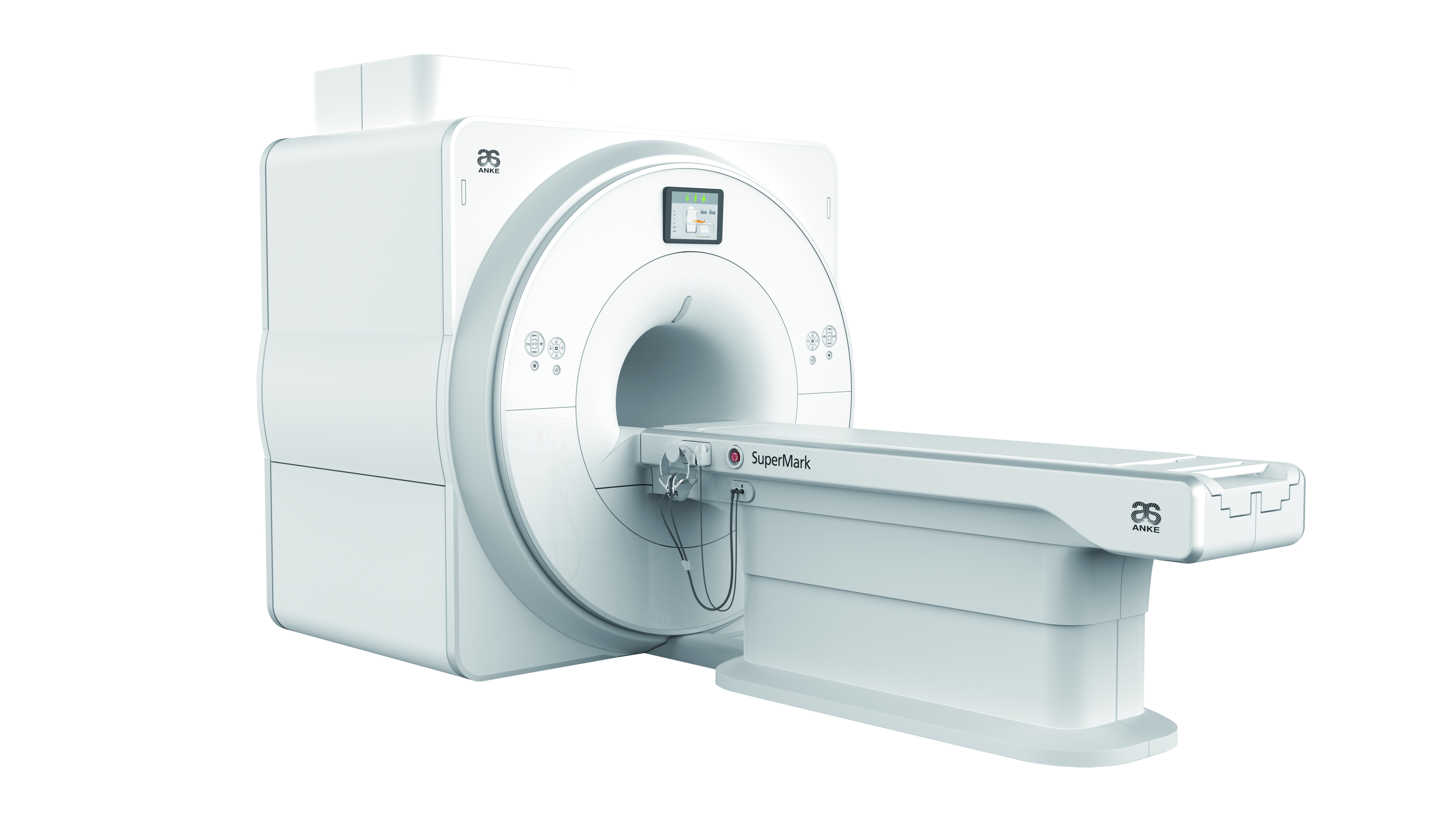
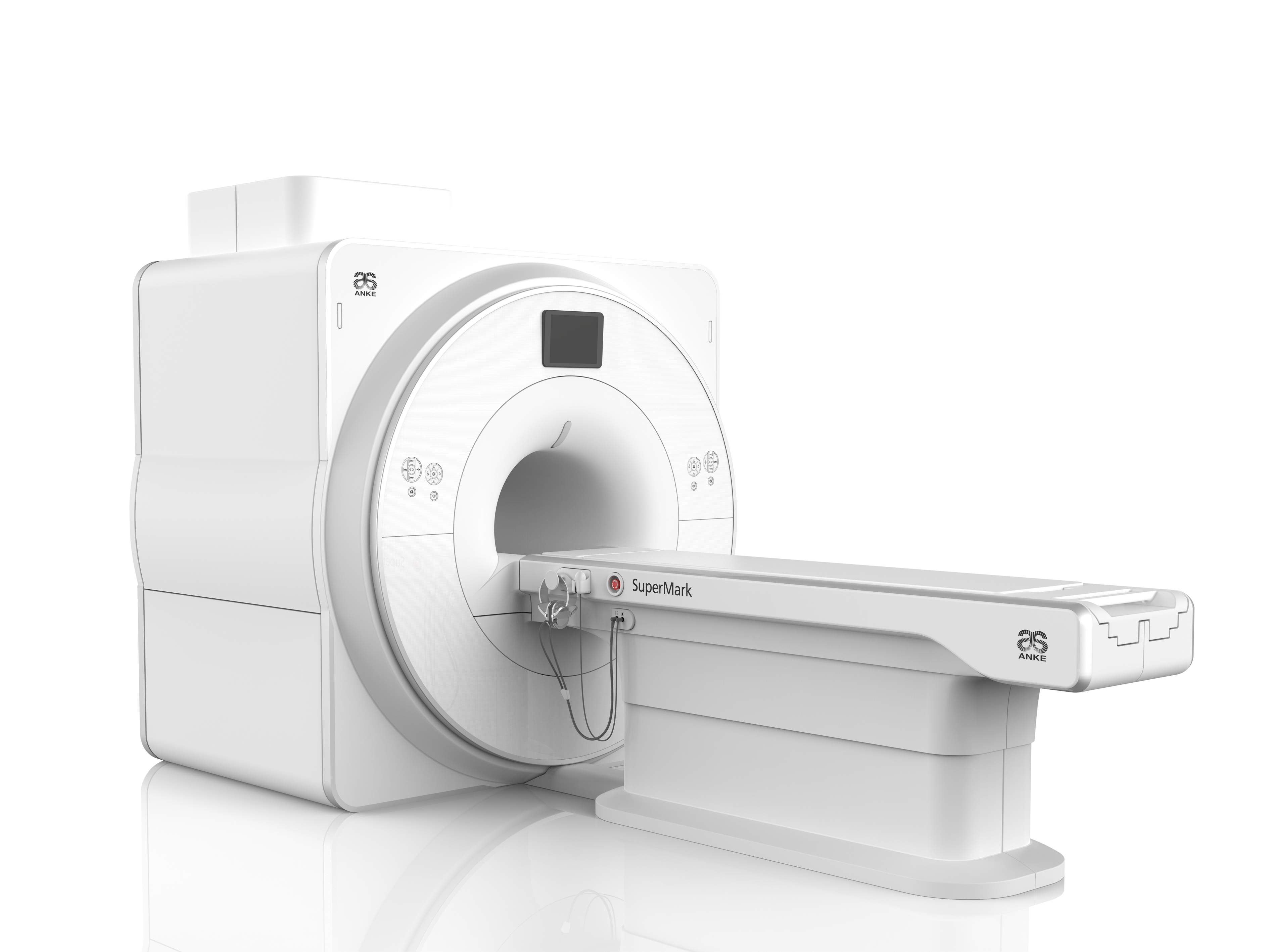
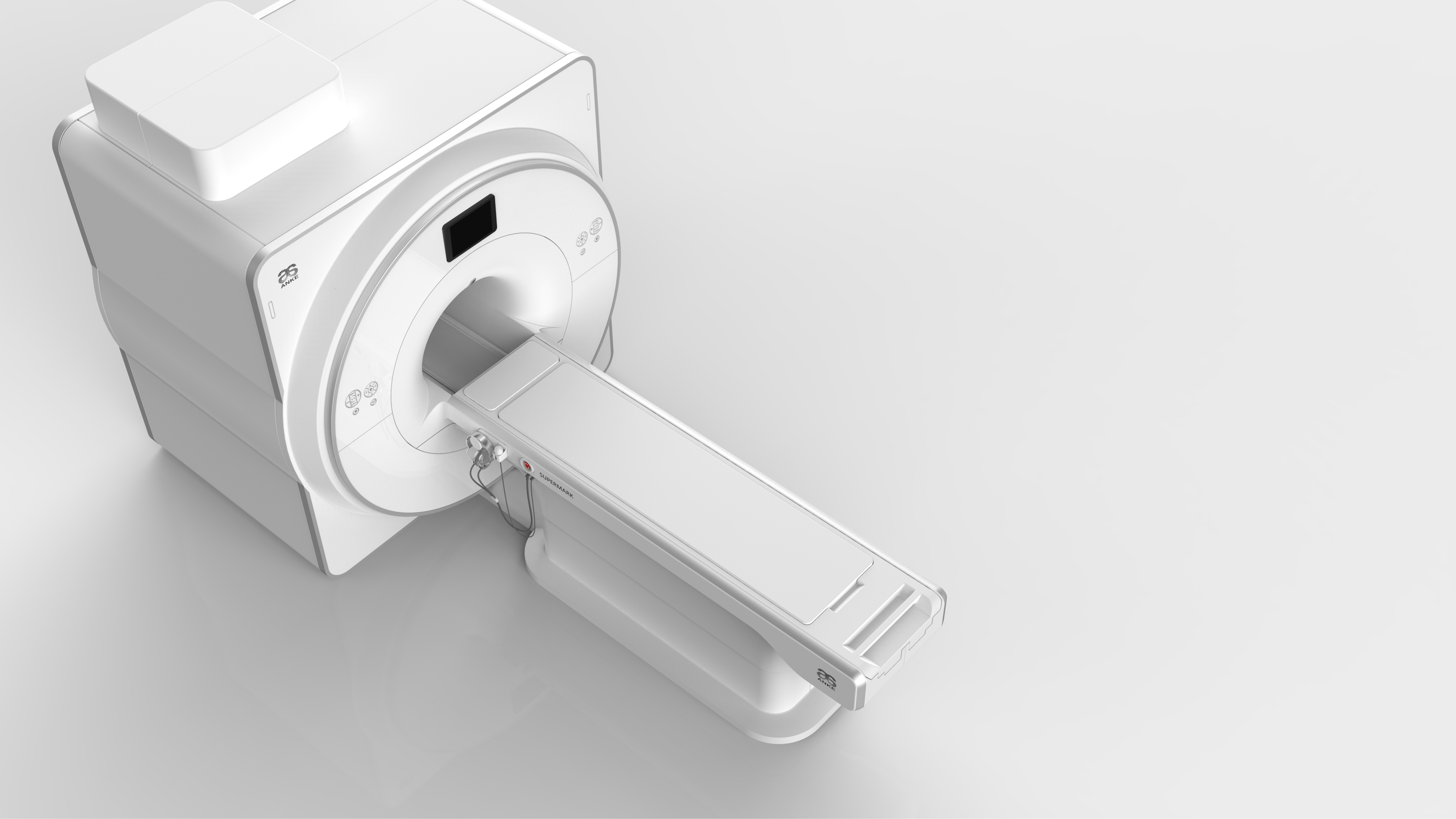
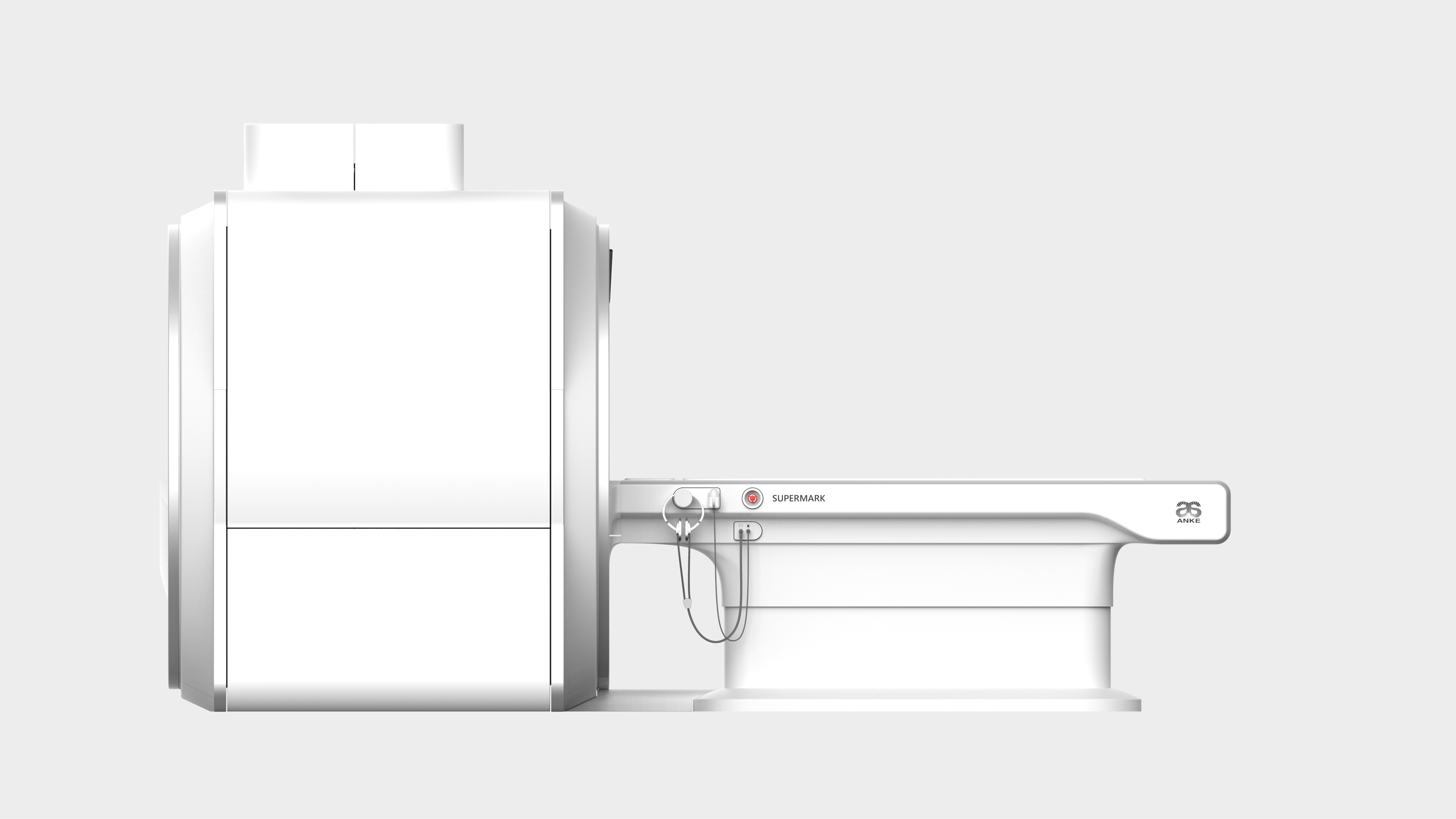
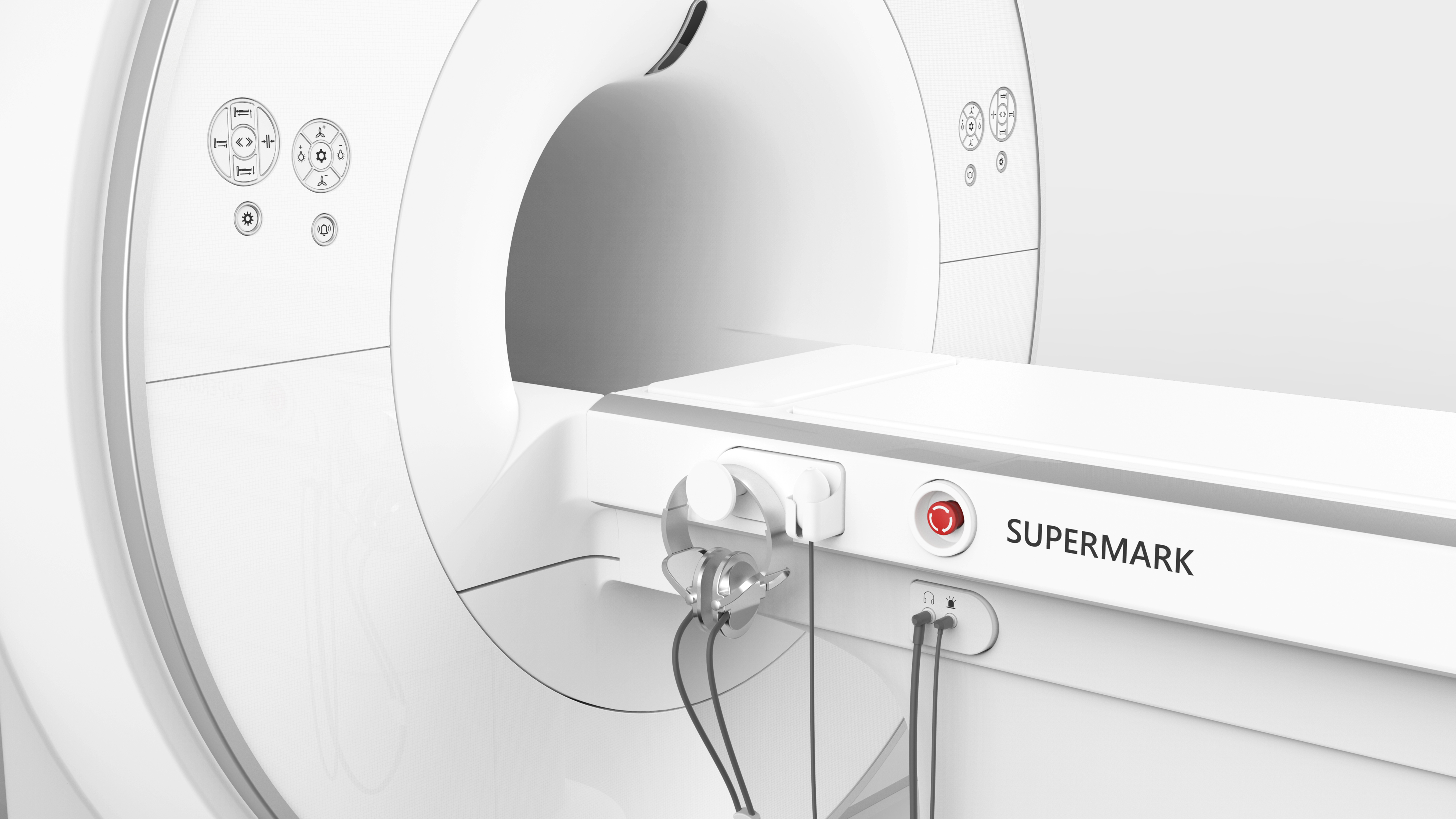
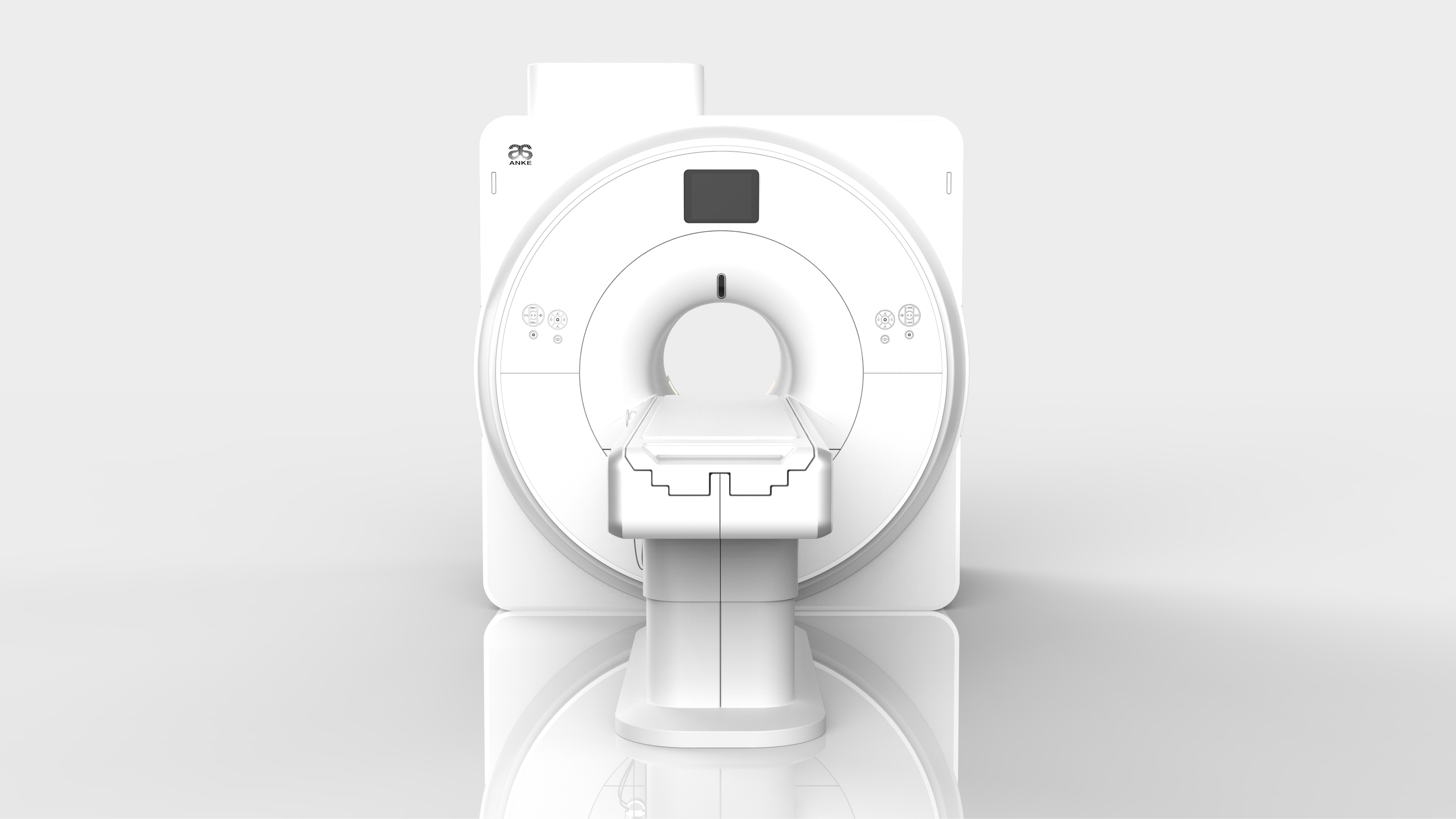
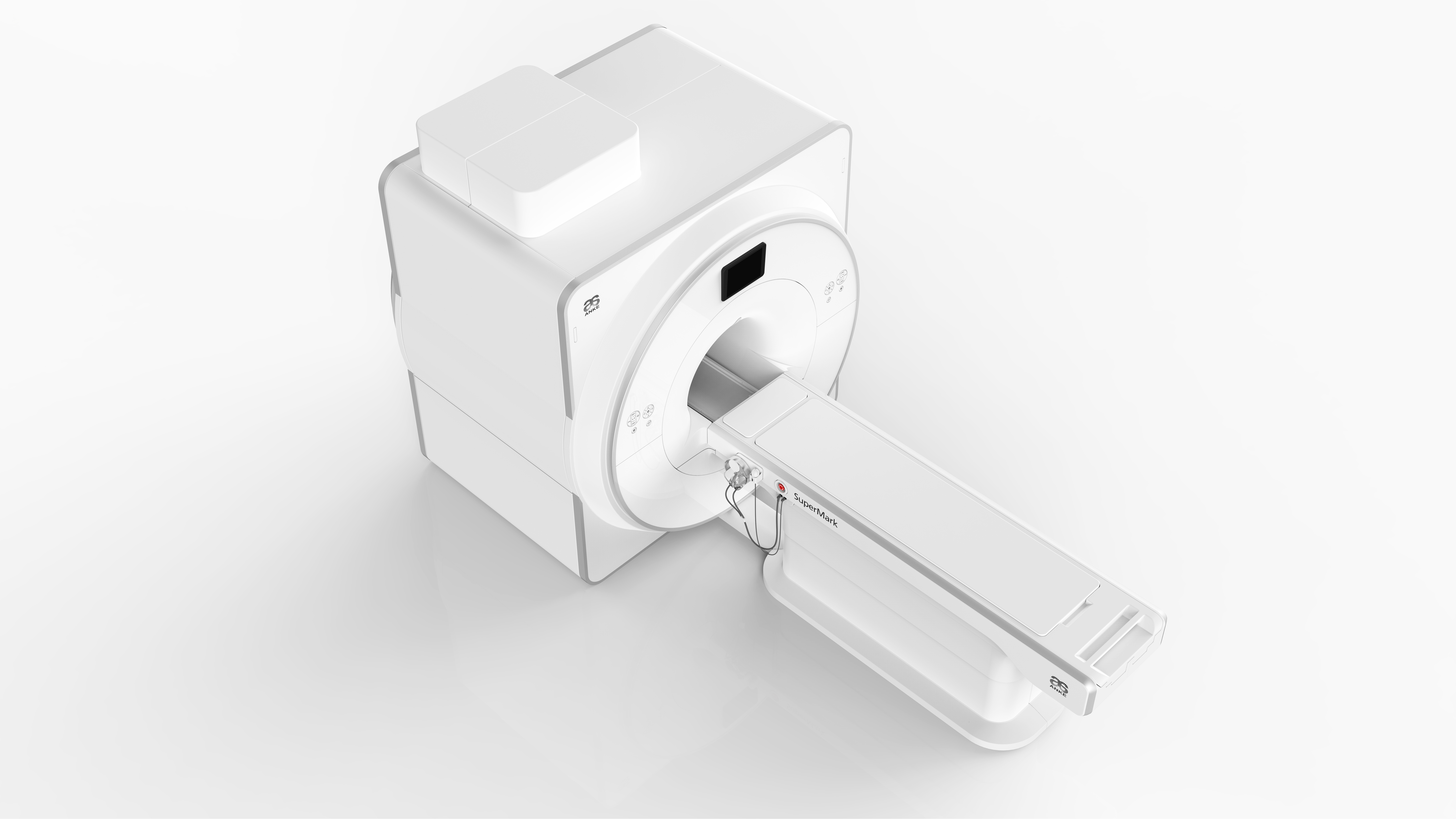
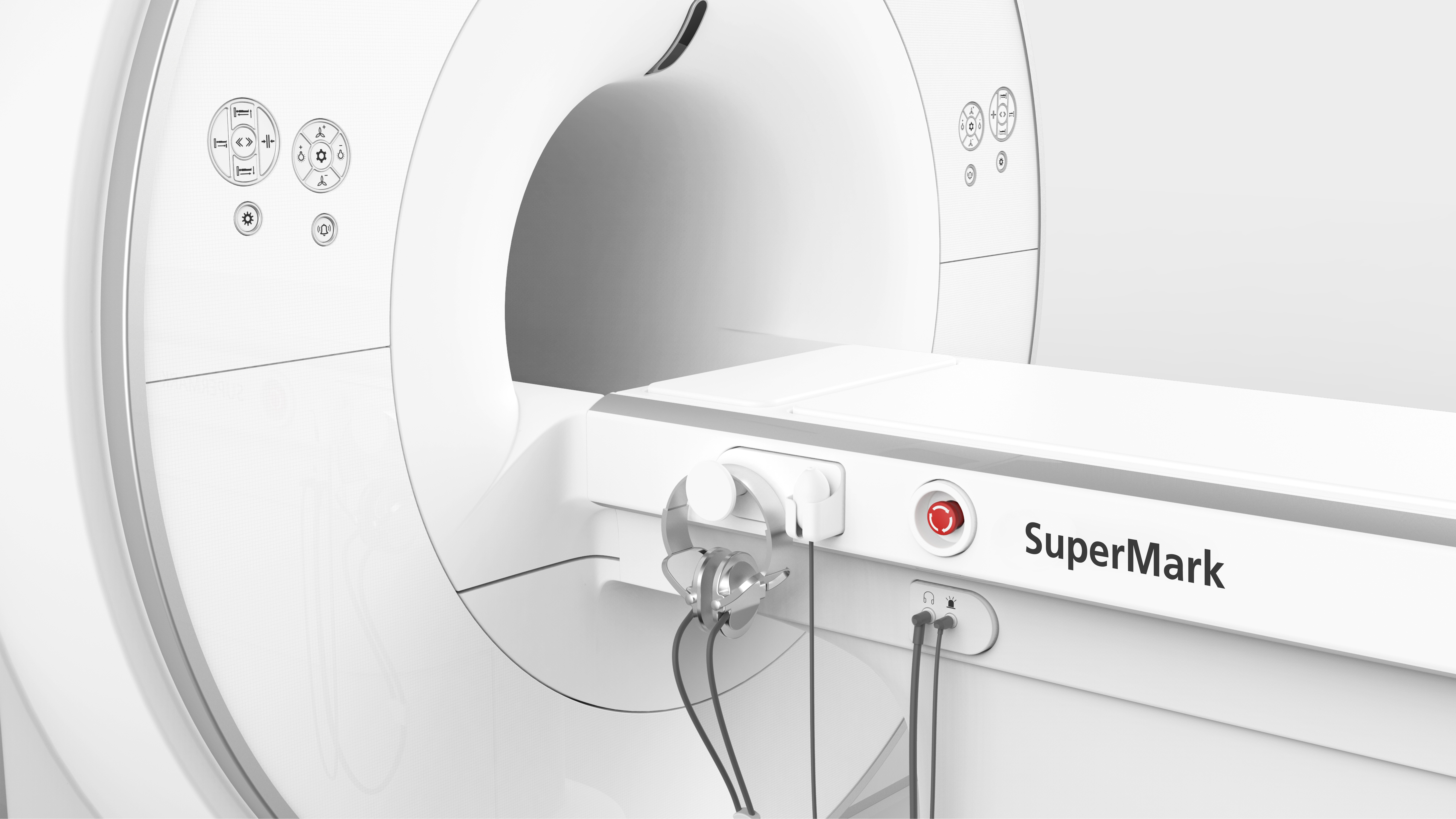
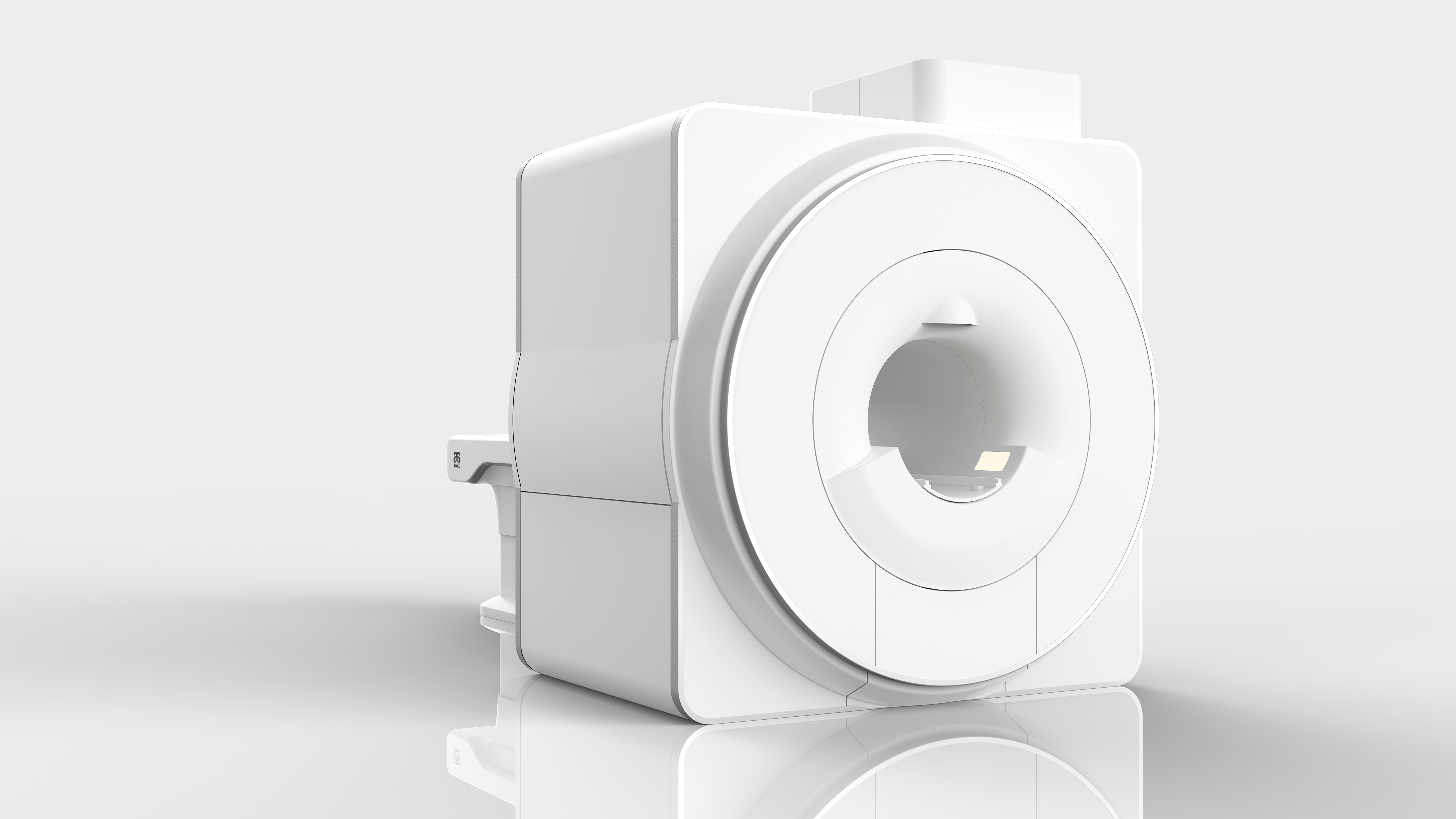
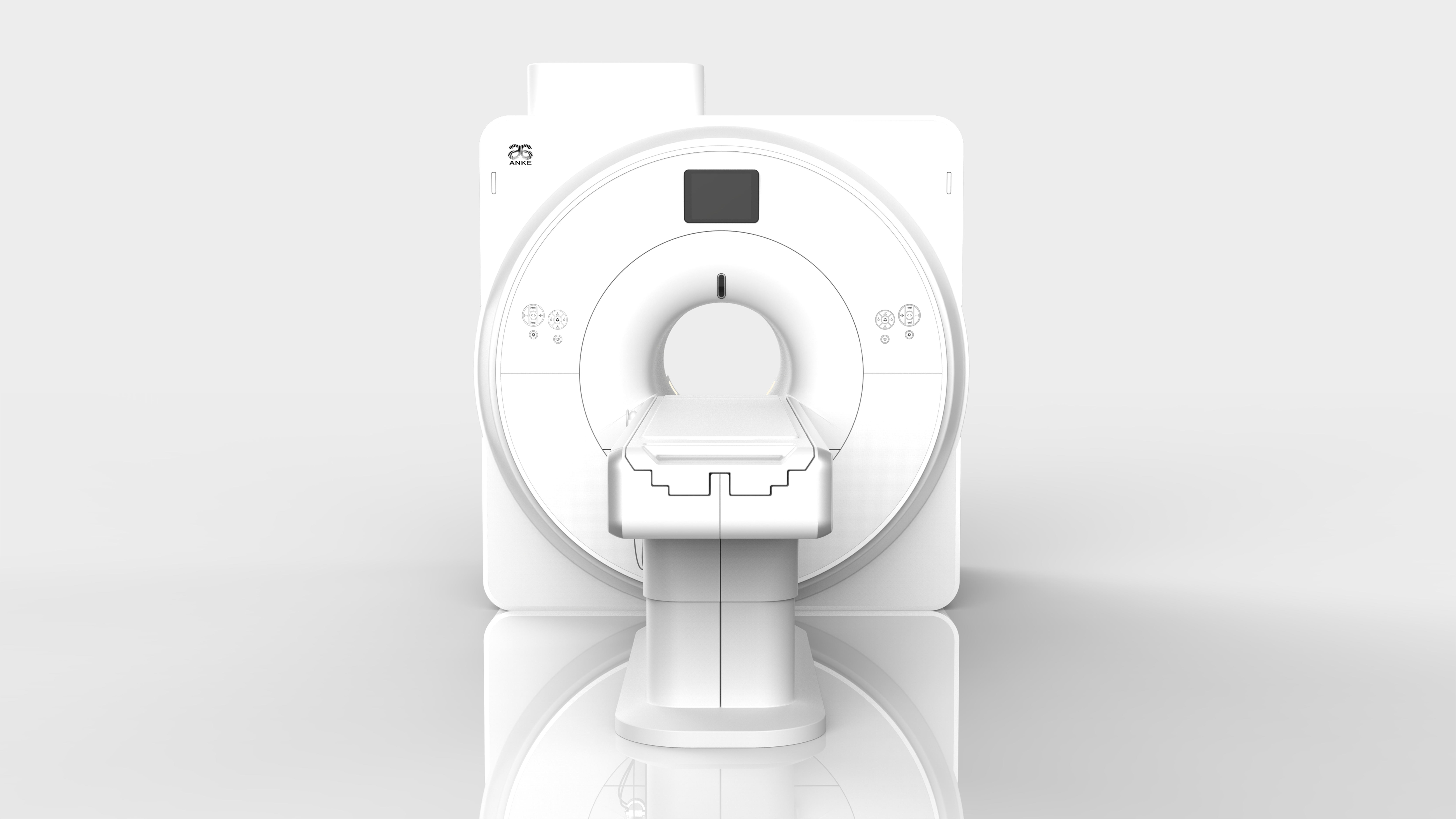
SuperMark 1.5T provides not only conventional pulse sequences and basic clinical applications, but also advanced functional applications. It adopts brand new APEX operating system which ensures easy operation and fast diagnosis.
Technical advantages |
- Short bore magnet with zero liquid helium “0” boil-off technology;
- Powerful and high linearity gradient system, Soft sound decreases the noise of scanning;
- High efficiency RF system combined with Dual-engine parallel acquisition technology;
- Multi-channel phased array receiving coil with intelligent identification;
- AI combined platform provide more comfortable operating experience;
- Complete clinical application package to meet all your clinical needs.
Short bore magnet |
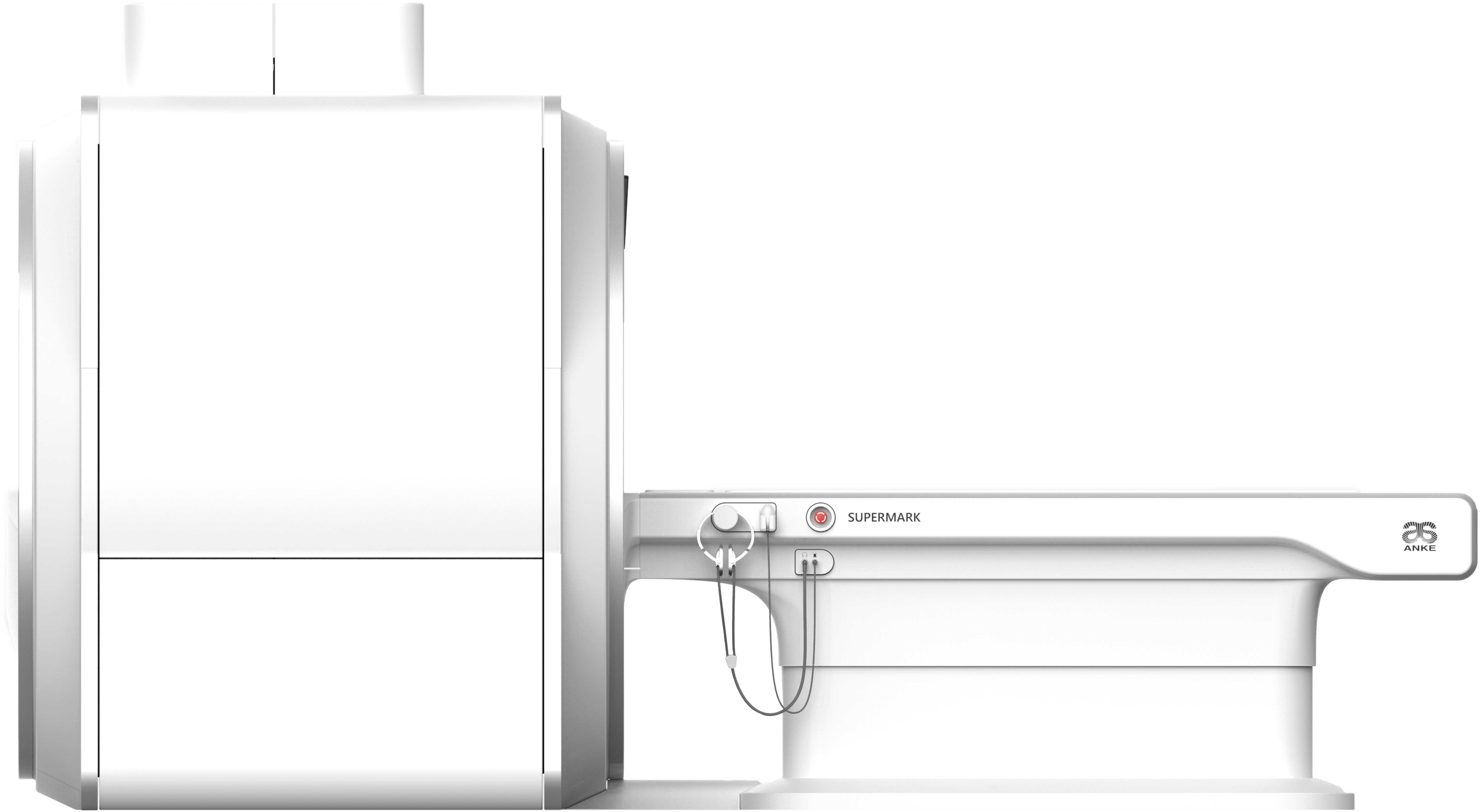
Traditional long magnet has a very long aperture, which seriously affects the comfort of patient examination. SuperMark 1.5T system is equipped with an optimally designed short bore superconducting magnet, the length of magnet is only 157cm, it can greatly improve the patient examination experience.
ADS technology |
Homogeneity of the main magnetic field is the basis of MR imaging. A good magnetic field homogeneity helps to realize a wide range of abdominal scanning and off-center joints scanning. In particular, fat suppression imaging has a high requirement for the homogeneity of magnetic field.
ANKE provides unique ADS (Automatic 3D dynamic shimming) technology, which automatically performs 3D dynamic shimming according to the tissue and structural characteristics of different parts of human body during scanning. This can greatly reduce problems such as image distortion and failure of fat suppression caused by local magnetic field inhomogeneities.
Liquid helium “0” boil-off |
Ultra-low liquid helium capacity design, only 700L (100% fill in) liquid helium is required to ensure a stable running of the magnet.
Equipped with 4K cold head and liquid helium “0” boil-off technology, which ensure stably running for more than 5 years without liquid helium fill in.
Safe and reliable |
With 24-hour unattended function, it can access the cloud server, monitor the magnet status online, and receive early warning information in real time to avoid major losses.
| Magnet specifications | |
| Magnet type | Superconducting |
| Magnetic field strength | 1.5T ± 1% |
| Magnet length | 157cm |
| Magnet bore diameter | 605mm±5mm |
| Weight (including 100% liquid helium) | 3800kg |
| Liquid helium volume (100% liquid helium) | 700L |
| Liquid refilling interval | ≥ 5 years |
| Magnetic field homogeneity | ≤ 0.170ppm @40cm DSV (VRSM) |
General features |
- Water-cooled gradient amplifier and coil, ensure the whole system working in maximum performance.
- Eddy-“0” technology, through the combination of hardware and software, the eddy current is compensated to the maximum extent, ensure excellent imaging quality.
- Ultrafast solid-state technology with very low switching losses.
- Excellent linearity, easy to achieve a wide range of scanning.
| Powerful output | |
| Max. amplitude (single axis) | 40mT/m |
| Max. Effective amplitude | 69mT/m |
| Max. slew rate (single axis) | 150T/m/s |
| Max. Effective slew rate | 259T/m/s |
| Min. rise time | 0.27ms |
| Duty cycle | 100% |
| The technology of maximum gradient amplitude and slew rate to arrive at the same time is provided. | |
Soft sound technology |
Structurally stable magnet and gradient coil with new design of noise reduction material, these two parts are closely connected, and the force is balanced in all directions, which can effectively reduce the vibration in the scanning process, thus reducing the noise.
The optimized sequence parameters can effectively avoid the resonance of the gradient coil, on the premise of ensuring image quality and scanning time, the noise generated by scanning is greatly reduced.
RF (Radio Frequency) system
General features |
- High efficiency RF system, equipped with new and unique technology, reducing electrical noise and increasing signal detection.
- Real-time RF energy monitoring technology, include short-term and long-term accumulation monitoring.
- 16 channels RF receiving platform, high dynamic range without adjustments.
RF (Radio Frequency) coils
Integrated body coil |
Transmitting & Receiving integrated Body Coil for whole-body coverage scan, can be seamlessly connected with the phased array coils, greatly improve SNR and uniformity of images.
Phased array coils |
The phased array coils are designed for high image quality. No-tune high element coils increase SNR and reduce examination times.
Head/Neck coil – 16 |
||
| Channels | 16 | |
| Dimensions (L×W×H) | 43cm×33cm×32cm | |
| Weight | 5.0kg | |
| Applications | • Head imaging
• Neck imaging • C-spine imaging • Head & neck MR angiography • Head & neck combined imaging • TMJ (temporomandibular joints) imaging |
|
Body coil – 16 |
||
| Channels | 16 | |
| Dimensions (L×W×H) | 55cm×46cm×3.5cm | |
| Weight | 4.6kg | |
| Applications | • Spine imaging
• Thorax imaging • Abdomen imaging • Pelvis imaging • Hip imaging • Abdomen MR angiography |
|
Knee coil – 8 |
||
| Channels | 8 | |
| Dimensions (L×W×H) | 42cm×27cm×27cm | |
| Weight | 4.9kg | |
| Applications | • High resolution knee imaging
• Lower extremities joint imaging |
|
Large flex coil – 4 |
||
| Channels | 4 | |
| Dimensions (L×W×H) | 51cm×23cm×2.7cm | |
| Weight | 1.4kg | |
| Applications | Imaging of large regions, such as medium to large shoulder, hip, and knee. | |
Head coil – 8 (Optional) |
||
| Channels | 8 | |
| Dimensions (L×W×H) | 32cm×40cm×31cm | |
| Weight | 4.3kg | |
| Applications | • Head imaging
• Head MR angiography • TMJ (temporomandibular joints) imaging |
|
Neck coil – 8 (Optional) |
||
| Channels | 8 | |
| Dimensions (L×W×H) | 70.5cm×42cm×31.7cm | |
| Weight | 7kg | |
| Applications | • Neck imaging
• C-spine & T-spine imaging • Neck MR angiography |
|
Hand/Wrist coil – 8 (Optional) |
||
| Channels | 8 | |
| Dimensions (L×W×H) | 32cm×16cm×23cm | |
| Weight | 4.6kg | |
| Applications | High resolution hand and wrist imaging | |
Foot/Ankle coil – 8 (Optional) |
||
| Channels | 8 | |
| Dimensions (L×W×H) | 37cm×30cm×32cm | |
| Weight | 6.5kg | |
| Applications | High resolution foot and ankle imaging | |
Breast coil – 8 (Optional) |
||
| Channels | 8 | |
| Dimensions (L×W×H) | 50cm×45cm×24cm | |
| Weight | 6.5kg | |
| Applications | • High resolution breast imaging
• Simultaneous imaging of both breasts • Axillar imaging elements |
|
Shoulder coil – 4 (Optional) |
||
| Channels | 4 | |
| Dimensions (L×W×H) | 21cm×29cm×19cm | |
| Weight | 5kg | |
| Applications | • High resolution shoulder imaging
• Higher SNR and homogeneity |
|
Small flex coil – 4 (Optional) |
||
| Channels | 4 | |
| Dimensions (L×W×H) | 36cm×18cm×2.7cm | |
| Weight | 0.8kg | |
| Applications | Imaging of small regions, such as small to medium shoulder, wrist, elbow, and ankle. | |
CTL Spine coil – 14 (Optional) |
||
| Channels | 14 | |
| Dimensions (L×W×H) | 112cm×42cm×30cm | |
| Weight | 9kg | |
| Applications | High resolution imaging of the whole spine | |
1) All the Optional coils are not included in the standard offer, please contact to ANKE for future information about technology and price.
Full soft-sound platform |
Combined with the advanced hardware design, Soft sound technology can be used for whole body scanning without image quality lose, which will provide a more comfortable scanning experience for patients. All the images bellow are collected by soft-sound sequence.
Dual-engine parallel acquisition technology |
Parallel acquisition is a technology to reduce the time of signal acquisition, in SuperMark 1.5T, we supply 2 kind of reconstruction method:
- SENSE (SENSitivity Encoding) technology based on Image domain.
- GRAPPA (GeneRalized Auto calibrating Partially Parallel Acquisition) technology based one K-space.
Parallel acquisition technology can fully satisfy your daily application needs and enhance the work efficiency.
AI technology for MRI imaging |
With the development of computer technology, Artificial Intelligence (AI) technology has been more and more widely used in some high-tech fields. Especially in the field of medical imaging, AI technology is also increasingly used in process optimization, image processing, and auxiliary diagnosis, etc. which can greatly improve work efficiency and reduce the workload of medical workers. As one of the top leading high-tech medical imaging manufacturers, ANKE has always been committed to the research on the application of AI technology in MRI.
- AI-Auto Localization technology
AI-Auto Localization technology uses AI to separate the tissues of the vertebral body, then find the corresponding intervertebral disc based on the vertebral body and place the cross-sectional positioning line parallel to the long axis of the intervertebral disc and perpendicular to the sagittal plane of the spine. This can greatly reduce the complexity of the operation, also, it can save time and avoid position and angle errors caused by manual operation.
- AI-Image Enhancement
Combined with fast scanning sequence, AI image enhancement technology can greatly shorten patient scanning time, improve patient comfort, and increase the number of patient scans in the hospital every day.
- AI motion correction
AI motion correction can reduce the image artifact by different data acquisition and reconstruction methods.
Multiple angiography imaging |
TOF (Time-of-Flight) 2D/3D based on the enhancement effect of blood flow, and is mainly used for the examination of cerebral blood vessels, neck blood vessels, and lower limb blood vessels.
CEMRA (Contrast Enhanced MR Angiography) 2D/3D for single step, dynamic, peripheral with the shortest TR and TE. The strong gradients make it possible to separate the arterial phase from the venous phase.
PCMRA (Phase Contrast MR Angiography) can be used for venous or sinus scanning, cerebrospinal fluid or blood flow, flow, flow direction analysis, etc.
ASL-MRA (Arterial Spin Labeling MR angiography) is a non-contrast MRA imaging function based on SSFP sequence. This function can be used to produce images of portal veins, renal arteries, and upper and lower extremity arteries.
MTC (Magnetization Transfer Contrast) technology and TONE (Tilted Optimized Non-saturation Excitation) pulse to improved Contrast to Noise Ratio of images.
High-definition diffusion imaging |
DWI (Diffusion Weighted Imaging) is to show Brownian motion of water molecules by applying diffusion sensitive gradient on the basis of EPI (Echo Plane Imaging) sequence.
In SuperMark 1.5T system, we supply EPI (Echo Planar Imaging) with Single Shot and Multi Shot technology for high-definition diffusion weighted imaging. In one scanning, you can obtain 4 images of DWI: DWI images with high b-value, DWI images with 0 b-value, ADC-map, eADC-map.
DWI is a necessary function for the examination cerebral infarction, brain tumor, malignant lesions (body) and bone tumor, etc.
DTI (Diffusion Tensor Imaging)1) is a more accurate description of the anisotropy of water molecular motion, with multiple diffusion-weightings and up to 256 directions for generating data sets for diffusion tensor imaging.
2) Optional functions. To obtain more accurate image information, the Diffusion tensor imaging (DTI) analysis software (optional) and other accessories are required.
Susceptibility weighted imaging (SWI) |
SWI (Susceptibility weighted imaging) is using the magnetic sensitive characteristics of deoxyhemoglobin, to get the image of veins, bleeding and non-heme iron deposits.
Amide Proton Transfer Imaging |
APT (Amide Proton Transfer) imaging is a noninvasive imaging technique based on chemical exchange saturation transfer technology.
APT is a reflection of protein distribution in MRI which can be used to assess protein expression of tumor, stroke and other brain diseases, and provides important information for diagnosis and treatment.
Perfusion weighted imaging2) |
PWI (Perfusion Weighted Imaging)3) is observing the changes in cerebral hemodynamics by EPI technology.
Inline Perfusion helps to streamline the clinical workflow by automating post-processing perfusion data during data acquisition. Neuro Perfusion measures perfusion deficits and assist in the diagnosis and grading of e.g. vascular deficiencies and brain tumors.
Dynamic contrast enhancement imaging |
DCE (Dynamic Contrast Enhanced) is a noninvasive imaging method to detect and evaluate the vascular status of tumors or other tissues by injecting contrast agents.
FAST 3D – DCE (Dynamic Contrast Enhancement) imaging with full abdominal coverage scan, can easily to capture the process of lesion signal enhancement, help you analyze the lesion more accurately.
3) Optional functions. To obtain more accurate image information, the MRI perfusion analysis software (optional) and other accessories are required.
Multiple fat and water imaging |
SuperMark 1.5T provide multiple precise fat and water imaging technologies.
STIR (Short Time Inversion Recovery)
FS (Fat Saturation) technology based on frequency selective RF pulses with 2 selectable modes: weak, strong
SPAIR (Spectral Adiabatic Inversion Recovery), combined with frequency selective inversion pulse to obtain a high-quality fat suppression body imaging
WE (Water Excitation) technology, can show the cartilage structure clearly
DIXON (Water and Fat Separation) technology.
Installation
Power Requirements |
380VAC ± 10%, 50/60 Hz, ±1Hz, Connection value 80 kVA
Recommended dimension3) |
| Scanning room | 6.00m x 5.00m |
| Operation room | 3.00m x 3.00m |
| Equipment room | 3.00m x 4.00m |


